Journal: Nature / Year: 2024 Title: Design of customized coronavirus receptors. Authors: Peng Liu / Mei-Ling Huang / Hua Guo / Matthew McCallum / Jun-Yu Si / Yuan-Mei Chen / Chun-Li Wang / Xiao Yu / Lu-Lu Shi / Qing Xiong / Cheng-Bao Ma / John E Bowen / Fei Tong / Chen Liu / Ye- ...Authors: Peng Liu / Mei-Ling Huang / Hua Guo / Matthew McCallum / Jun-Yu Si / Yuan-Mei Chen / Chun-Li Wang / Xiao Yu / Lu-Lu Shi / Qing Xiong / Cheng-Bao Ma / John E Bowen / Fei Tong / Chen Liu / Ye-Hui Sun / Xiao Yang / Jing Chen / Ming Guo / Jing Li / Davide Corti / David Veesler / Zheng-Li Shi / Huan Yan / Abstract: Although coronaviruses use diverse receptors, the characterization of coronaviruses with unknown receptors has been impeded by a lack of infection models. Here we introduce a strategy to engineer ...Although coronaviruses use diverse receptors, the characterization of coronaviruses with unknown receptors has been impeded by a lack of infection models. Here we introduce a strategy to engineer functional customized viral receptors (CVRs). The modular design relies on building artificial receptor scaffolds comprising various modules and generating specific virus-binding domains. We identify key factors for CVRs to functionally mimic native receptors by facilitating spike proteolytic cleavage, membrane fusion, pseudovirus entry and propagation for various coronaviruses. We delineate functional SARS-CoV-2 spike receptor-binding sites for CVR design and reveal the mechanism of cell entry promoted by the N-terminal domain-targeting S2L20-CVR. We generated CVR-expressing cells for 12 representative coronaviruses from 6 subgenera, most of which lack known receptors, and show that a pan-sarbecovirus CVR supports propagation of a propagation-competent HKU3 pseudovirus and of authentic RsHuB2019A. Using an HKU5-specific CVR, we successfully rescued wild-type and ZsGreen-HiBiT-incorporated HKU5-1 (LMH03f) and isolated a HKU5 strain from bat samples. Our study demonstrates the potential of the CVR strategy for establishing native receptor-independent infection models, providing a tool for studying viruses that lack known susceptible target cells.
In the structure databanks used in Yorodumi, some data are registered as the other names, "COVID-19 virus" and "2019-nCoV". Here are the details of the virus and the list of structure data.
Jan 31, 2019. EMDB accession codes are about to change! (news from PDBe EMDB page)
EMDB accession codes are about to change! (news from PDBe EMDB page)
The allocation of 4 digits for EMDB accession codes will soon come to an end. Whilst these codes will remain in use, new EMDB accession codes will include an additional digit and will expand incrementally as the available range of codes is exhausted. The current 4-digit format prefixed with “EMD-” (i.e. EMD-XXXX) will advance to a 5-digit format (i.e. EMD-XXXXX), and so on. It is currently estimated that the 4-digit codes will be depleted around Spring 2019, at which point the 5-digit format will come into force.
The EM Navigator/Yorodumi systems omit the EMD- prefix.
Related info.:Q: What is EMD? / ID/Accession-code notation in Yorodumi/EM Navigator
Yorodumi is a browser for structure data from EMDB, PDB, SASBDB, etc.
This page is also the successor to EM Navigator detail page, and also detail information page/front-end page for Omokage search.
The word "yorodu" (or yorozu) is an old Japanese word meaning "ten thousand". "mi" (miru) is to see.
Related info.:EMDB / PDB / SASBDB / Comparison of 3 databanks / Yorodumi Search / Aug 31, 2016. New EM Navigator & Yorodumi / Yorodumi Papers / Jmol/JSmol / Function and homology information / Changes in new EM Navigator and Yorodumi
 Movie
Movie Controller


 Open data
 Basic information
 Map data
 Sample
 Keywords
 Function and homology information
 Homo sapiens (human) /
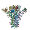
 Severe acute respiratory syndrome coronavirus 2
 Authors
 United States, 1 items
  Citation

 Structure visualization
 Downloads & links
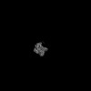
 emd_45174.png
 http://ftp.pdbj.org/pub/emdb/structures/EMD-45174
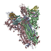


 Z (Sec.)
 Y (Row.)
 X (Col.)


 Sample components
 Processing
 Electron microscopy
 FIELD EMISSION GUN