 Movie
Movie Controller


+ Open data

- Basic information
| Entry | Database: PDB / ID: 9c1u | |||||||||
|---|---|---|---|---|---|---|---|---|---|---|
| Title | Cryo-EM Structure of a Tm1C Fibril | |||||||||
 Components Components | Tropomyosin 1 I/C | |||||||||
 Keywords Keywords | PROTEIN FIBRIL | |||||||||
| Function / homology | Tropomyosin / Tropomyosin / GH09289p Function and homology information Function and homology information | |||||||||
| Biological species |  | |||||||||
| Method | ELECTRON MICROSCOPY / helical reconstruction / cryo EM / Resolution: 2.31 Å | |||||||||
 Authors Authors | Fonda, B.D. / Kato, M. / Li, Y. / Murray, D.T. | |||||||||
| Funding support |  United States, 2items United States, 2items
| |||||||||
 Citation Citation | Journal: Protein Sci / Year: 2024 Title: Cryo-EM and solid state NMR together provide a more comprehensive structural investigation of protein fibrils. Authors: Blake D Fonda / Masato Kato / Yang Li / Dylan T Murray / Abstract: The tropomyosin 1 isoform I/C C-terminal domain (Tm1-LC) fibril structure is studied jointly with cryogenic electron microscopy (cryo-EM) and solid state nuclear magnetic resonance (NMR). This study ...The tropomyosin 1 isoform I/C C-terminal domain (Tm1-LC) fibril structure is studied jointly with cryogenic electron microscopy (cryo-EM) and solid state nuclear magnetic resonance (NMR). This study demonstrates the complementary nature of these two structural biology techniques. Chemical shift assignments from solid state NMR are used to determine the secondary structure at the level of individual amino acids, which is faithfully seen in cryo-EM reconstructions. Additionally, solid state NMR demonstrates that the region not observed in the reconstructed cryo-EM density is primarily in a highly mobile random coil conformation rather than adopting multiple rigid conformations. Overall, this study illustrates the benefit of investigations combining cryo-EM and solid state NMR to investigate protein fibril structure. | |||||||||
| History |
|
- Structure visualization
| Structure viewer | Molecule: MolmilJmol/JSmol |
|---|
- Downloads & links
-Download
| PDBx/mmCIF format | 9c1u.cif.gz | 71.9 KB | Display | PDBx/mmCIF format |
|---|---|---|---|---|
| PDB format | pdb9c1u.ent.gz | 51.5 KB | Display | PDB format |
| PDBx/mmJSON format | 9c1u.json.gz | Tree view | PDBx/mmJSON format | |
| Others |  Other downloads Other downloads |
-Validation report
| Arichive directory | https://data.pdbj.org/pub/pdb/validation_reports/c1/9c1uftp://data.pdbj.org/pub/pdb/validation_reports/c1/9c1u | HTTPS FTP |
|---|
-Related structure data
| Related structure data |  45130MC M: map data used to model this data C: citing same article ( |
|---|---|
| Similar structure data |
-Links
 PDBj
PDBj- Assembly
| Deposited unit | 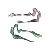
|
|---|---|
| 1 |
|
-Components
| #1: Protein | Mass: 7420.535 Da / Num. of mol.: 12 / Fragment: C-terminal domain, residues 373-441 Source method: isolated from a genetically manipulated source Details: SY-tagged Tm1 I/C alternative isoform / Source: (gene. exp.) Gene: Tm1, 1305/10, 2299, BcDNA:GH09289, BcDNA:LD37158, BcDNA:SD21996, chr3R:11122272..11122408, cTM, cTm, cTmII, Dm Tm1, Dm TmH33, Dm TmH34, Dmel\CG4898, DmTm1, l(3)02299, l(3)S130510, l(3)s2958, ...Gene: Tm1, 1305/10, 2299, BcDNA:GH09289, BcDNA:LD37158, BcDNA:SD21996, chr3R:11122272..11122408, cTM, cTm, cTmII, Dm Tm1, Dm TmH33, Dm TmH34, Dmel\CG4898, DmTm1, l(3)02299, l(3)S130510, l(3)s2958, mTmII, PmI, region 3, TM, Tm, TM1, tm1, TmH, TmH-33, TmH-34, TmH33, TmH34, TMII, TmII, tmII, Tmr33, Tmr34, TnH, TnH-33, TnH-34, tropomyosin, CG4898, Dmel_CG4898, Production host:  Has protein modification | N | |
|---|
-Experimental details
-Experiment
| Experiment | Method: ELECTRON MICROSCOPY |
|---|---|
| EM experiment | Aggregation state: HELICAL ARRAY / 3D reconstruction method: helical reconstruction |
- Sample preparation
| Component | Name: Tropomyosin 1 I/C C-terminal Domain / Type: ORGANELLE OR CELLULAR COMPONENT / Entity ID: all / Source: RECOMBINANT | ||||||||||||||||||||||||
|---|---|---|---|---|---|---|---|---|---|---|---|---|---|---|---|---|---|---|---|---|---|---|---|---|---|
| Molecular weight | Value: 7.41557 kDa/nm / Experimental value: NO | ||||||||||||||||||||||||
| Source (natural) | Organism: | ||||||||||||||||||||||||
| Source (recombinant) | Organism: | ||||||||||||||||||||||||
| Buffer solution | pH: 7.5 | ||||||||||||||||||||||||
| Buffer component |
| ||||||||||||||||||||||||
| Specimen | Conc.: 1 mg/ml / Embedding applied: NO / Shadowing applied: NO / Staining applied: NO / Vitrification applied: YES | ||||||||||||||||||||||||
| Specimen support | Grid material: COPPER / Grid mesh size: 300 divisions/in. / Grid type: Quantifoil R1.2/1.3 | ||||||||||||||||||||||||
| Vitrification | Instrument: FEI VITROBOT MARK IV / Cryogen name: ETHANE / Humidity: 95 % / Chamber temperature: 277.15 K |
- Electron microscopy imaging
| Experimental equipment |  Model: Titan Krios / Image courtesy: FEI Company |
|---|---|
| Microscopy | Model: FEI TITAN KRIOS |
| Electron gun | Electron source:  FIELD EMISSION GUN / Accelerating voltage: 300 kV / Illumination mode: FLOOD BEAM FIELD EMISSION GUN / Accelerating voltage: 300 kV / Illumination mode: FLOOD BEAM |
| Electron lens | Mode: BRIGHT FIELD / Nominal defocus max: 2400 nm / Nominal defocus min: 1000 nm |
| Image recording | Electron dose: 52 e/Å2 / Film or detector model: GATAN K3 (6k x 4k) |
- Processing
| EM software |
| ||||||||||||||||||||||||
|---|---|---|---|---|---|---|---|---|---|---|---|---|---|---|---|---|---|---|---|---|---|---|---|---|---|
| CTF correction | Type: PHASE FLIPPING AND AMPLITUDE CORRECTION | ||||||||||||||||||||||||
| Helical symmerty | Angular rotation/subunit: 2.58 ° / Axial rise/subunit: 4.69 Å / Axial symmetry: C1 | ||||||||||||||||||||||||
| 3D reconstruction | Resolution: 2.31 Å / Resolution method: FSC 0.143 CUT-OFF / Num. of particles: 17145 / Num. of class averages: 1 / Symmetry type: HELICAL | ||||||||||||||||||||||||
| Refine LS restraints |
|
