 Movie
Movie Controller

[English] 日本語
 Yorodumi
- PDB-9c0n: FphI, Staphylococcus aureus fluorophosphonate-binding serine hydr... -
+ Open data

- Basic information
| Entry | Database: PDB / ID: 9c0n | ||||||
|---|---|---|---|---|---|---|---|
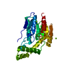
| Title | FphI, Staphylococcus aureus fluorophosphonate-binding serine hydrolases I, apo form at room temperature | ||||||
 Components Components | Alpha/beta fold hydrolase | ||||||
 Keywords Keywords | HYDROLASE / FphI / Staphylococcus aureus / S. aureus / fluorophosphonate-binding / serine hydrolases / lipase / room temperature / humidity / humidifier | ||||||
| Function / homology | Esterase/lipase / : / Serine aminopeptidase, S33 / carboxylesterase / Serine aminopeptidase, S33 / carboxylesterase activity / Alpha/Beta hydrolase fold / Alpha/beta fold hydrolase Function and homology information Function and homology information | ||||||
| Biological species |  Staphylococcus aureus USA300-CA-263 (bacteria) Staphylococcus aureus USA300-CA-263 (bacteria) | ||||||
| Method |  X-RAY DIFFRACTION / SYNCHROTRON / MOLECULAR REPLACEMENT / Resolution: 1.7 Å X-RAY DIFFRACTION / SYNCHROTRON / MOLECULAR REPLACEMENT / Resolution: 1.7 Å | ||||||
 Authors Authors | Fellner, M. / Randall, G.T. | ||||||
| Funding support | 1items
| ||||||
 Citation Citation | Journal: Proteins / Year: 2025 Title: Similar but Distinct-Biochemical Characterization of the Staphylococcus aureus Serine Hydrolases FphH and FphI. Authors: Fellner, M. / Randall, G. / Bitac, I.R.C.G. / Warrender, A.K. / Sethi, A. / Jelinek, R. / Kass, I. | ||||||
| History |
|
- Structure visualization
| Structure viewer | Molecule: MolmilJmol/JSmol |
|---|
- Downloads & links
-Download
| PDBx/mmCIF format | 9c0n.cif.gz | 162.2 KB | Display | PDBx/mmCIF format |
|---|---|---|---|---|
| PDB format | pdb9c0n.ent.gz | 129.4 KB | Display | PDB format |
| PDBx/mmJSON format | 9c0n.json.gz | Tree view | PDBx/mmJSON format | |
| Others |  Other downloads Other downloads |
-Validation report
| Arichive directory | https://data.pdbj.org/pub/pdb/validation_reports/c0/9c0nftp://data.pdbj.org/pub/pdb/validation_reports/c0/9c0n | HTTPS FTP |
|---|
-Related structure data


-Links
 PDBj
PDBj- Assembly
| Deposited unit | 
| ||||||||
|---|---|---|---|---|---|---|---|---|---|
| 1 |
| ||||||||
| Unit cell |
|
-Components
| #1: Protein | Mass: 27686.197 Da / Num. of mol.: 1 / Mutation: N-terminal GPG from expression tag Source method: isolated from a genetically manipulated source Source: (gene. exp.) Staphylococcus aureus USA300-CA-263 (bacteria)Gene: est_2, est_1, BN1321_100022, C7M54_03125, DD547_00417, EP54_01625, EQ90_13375, FAF17_11905, GO814_02325, GO941_12735, GO942_04080, GQX37_00145, HMPREF3211_01442, NCTC10702_00834, NCTC13131_ ...Gene: est_2, est_1, BN1321_100022, C7M54_03125, DD547_00417, EP54_01625, EQ90_13375, FAF17_11905, GO814_02325, GO941_12735, GO942_04080, GQX37_00145, HMPREF3211_01442, NCTC10702_00834, NCTC13131_05910, SAMEA2078260_02464, SAMEA2078588_01365, SAMEA2080344_01425, SAMEA2081063_02567, SAMEA4008575_01596, SAMEA70146418_02725 Production host: | ||||||
|---|---|---|---|---|---|---|---|
| #2: Chemical | ChemComp-CA /   Mass: 40.078 Da / Num. of mol.: 4 / Source method: obtained synthetically / Formula: Ca Mass: 40.078 Da / Num. of mol.: 4 / Source method: obtained synthetically / Formula: Ca#3: Water | ChemComp-HOH / |  Mass: 18.015 Da / Num. of mol.: 119 / Source method: isolated from a natural source / Formula: H2O Mass: 18.015 Da / Num. of mol.: 119 / Source method: isolated from a natural source / Formula: H2OHas ligand of interest | N | Has protein modification | N | |
-Experimental details
-Experiment
| Experiment | Method: X-RAY DIFFRACTION / Number of used crystals: 1 |
|---|
- Sample preparation
| Crystal | Density Matthews: 2.27 Å3/Da / Density % sol: 45.71 % |
|---|---|
| Crystal grow | Temperature: 289.15 K / Method: vapor diffusion, sitting drop / pH: 7 Details: 0.2 uL 10 mg/mL FphI (10 mM HEPES pH 7.5, 100 mM NaCl) were mixed with 0.2 uL of reservoir solution. Sitting drop reservoir contained 200mM Calcium chloride hexahydrate, 100mM HEPES pH 7.0, 20 % w/v PEG 6000. |
-Data collection
| Diffraction | Mean temperature: 295 K / Ambient temp details: Room temperature data collection / Serial crystal experiment: N |
|---|---|
| Diffraction source | Source: SYNCHROTRON / Site: SSRL  / Beamline: BL12-1 / Wavelength: 0.979 Å / Beamline: BL12-1 / Wavelength: 0.979 Å |
| Detector | Type: DECTRIS EIGER X 16M / Detector: PIXEL / Date: Mar 13, 2024 |
| Radiation | Protocol: SINGLE WAVELENGTH / Monochromatic (M) / Laue (L): M / Scattering type: x-ray |
| Radiation wavelength | Wavelength: 0.979 Å / Relative weight: 1 |
| Reflection | Resolution: 1.7→48.21 Å / Num. obs: 28252 / % possible obs: 99.7 % / Redundancy: 5.2 % / CC1/2: 1 / Rmerge(I) obs: 0.049 / Rpim(I) all: 0.024 / Rrim(I) all: 0.055 / Χ2: 1.01 / Net I/σ(I): 15.8 / Num. measured all: 146228 |
| Reflection shell | Resolution: 1.7→1.73 Å / % possible obs: 98.5 % / Redundancy: 4.8 % / Rmerge(I) obs: 1.127 / Num. measured all: 6850 / Num. unique obs: 1435 / CC1/2: 0.66 / Rpim(I) all: 0.559 / Rrim(I) all: 1.263 / Χ2: 0.94 / Net I/σ(I) obs: 1.3 |
- Processing
| Software |
| |||||||||||||||||||||||||||||||||||||||||||||||||||||||||||||||||||||||||||||||||||||||||||||||||||||||||||||||||||||||||||||||||||||||||||||||||||||||||||||||||||||||||||||||
|---|---|---|---|---|---|---|---|---|---|---|---|---|---|---|---|---|---|---|---|---|---|---|---|---|---|---|---|---|---|---|---|---|---|---|---|---|---|---|---|---|---|---|---|---|---|---|---|---|---|---|---|---|---|---|---|---|---|---|---|---|---|---|---|---|---|---|---|---|---|---|---|---|---|---|---|---|---|---|---|---|---|---|---|---|---|---|---|---|---|---|---|---|---|---|---|---|---|---|---|---|---|---|---|---|---|---|---|---|---|---|---|---|---|---|---|---|---|---|---|---|---|---|---|---|---|---|---|---|---|---|---|---|---|---|---|---|---|---|---|---|---|---|---|---|---|---|---|---|---|---|---|---|---|---|---|---|---|---|---|---|---|---|---|---|---|---|---|---|---|---|---|---|---|---|---|---|
| Refinement | Method to determine structure: MOLECULAR REPLACEMENT / Resolution: 1.7→40.06 Å / SU ML: 0.16 / Cross valid method: FREE R-VALUE / σ(F): 1.34 / Phase error: 18.21 / Stereochemistry target values: ML
| |||||||||||||||||||||||||||||||||||||||||||||||||||||||||||||||||||||||||||||||||||||||||||||||||||||||||||||||||||||||||||||||||||||||||||||||||||||||||||||||||||||||||||||||
| Solvent computation | Shrinkage radii: 0.9 Å / VDW probe radii: 1.1 Å / Solvent model: FLAT BULK SOLVENT MODEL | |||||||||||||||||||||||||||||||||||||||||||||||||||||||||||||||||||||||||||||||||||||||||||||||||||||||||||||||||||||||||||||||||||||||||||||||||||||||||||||||||||||||||||||||
| Refinement step | Cycle: LAST / Resolution: 1.7→40.06 Å
| |||||||||||||||||||||||||||||||||||||||||||||||||||||||||||||||||||||||||||||||||||||||||||||||||||||||||||||||||||||||||||||||||||||||||||||||||||||||||||||||||||||||||||||||
| Refine LS restraints |
| |||||||||||||||||||||||||||||||||||||||||||||||||||||||||||||||||||||||||||||||||||||||||||||||||||||||||||||||||||||||||||||||||||||||||||||||||||||||||||||||||||||||||||||||
| LS refinement shell |
| |||||||||||||||||||||||||||||||||||||||||||||||||||||||||||||||||||||||||||||||||||||||||||||||||||||||||||||||||||||||||||||||||||||||||||||||||||||||||||||||||||||||||||||||
| Refinement TLS params. | Method: refined / Refine-ID: X-RAY DIFFRACTION
| |||||||||||||||||||||||||||||||||||||||||||||||||||||||||||||||||||||||||||||||||||||||||||||||||||||||||||||||||||||||||||||||||||||||||||||||||||||||||||||||||||||||||||||||
| Refinement TLS group |
|
