 Movie
Movie Controller

[English] 日本語
 Yorodumi
- PDB-9c0l: FphH, Staphylococcus aureus fluorophosphonate-binding serine hydr... -
+ Open data

- Basic information
| Entry | Database: PDB / ID: 9c0l | ||||||
|---|---|---|---|---|---|---|---|
| Title | FphH, Staphylococcus aureus fluorophosphonate-binding serine hydrolases H, apo crystal form 2 | ||||||
 Components Components | Alpha/beta fold hydrolase | ||||||
 Keywords Keywords | HYDROLASE / FphH / Staphylococcus aureus / S. aureus / fluorophosphonate-binding / serine hydrolases / lipase | ||||||
| Function / homology | Esterase/lipase / : / Serine aminopeptidase, S33 / carboxylesterase / Serine aminopeptidase, S33 / carboxylesterase activity / Alpha/Beta hydrolase fold / Carboxylesterase Function and homology information Function and homology information | ||||||
| Biological species |  Staphylococcus aureus USA300-CA-263 (bacteria) Staphylococcus aureus USA300-CA-263 (bacteria) | ||||||
| Method |  X-RAY DIFFRACTION / SYNCHROTRON / MOLECULAR REPLACEMENT / Resolution: 1.79 Å X-RAY DIFFRACTION / SYNCHROTRON / MOLECULAR REPLACEMENT / Resolution: 1.79 Å | ||||||
 Authors Authors | Fellner, M. | ||||||
| Funding support |  New Zealand, 1items New Zealand, 1items
| ||||||
 Citation Citation | Journal: Proteins / Year: 2025 Title: Similar but Distinct-Biochemical Characterization of the Staphylococcus aureus Serine Hydrolases FphH and FphI. Authors: Fellner, M. / Randall, G. / Bitac, I.R.C.G. / Warrender, A.K. / Sethi, A. / Jelinek, R. / Kass, I. | ||||||
| History |
|
- Structure visualization
| Structure viewer | Molecule: MolmilJmol/JSmol |
|---|
- Downloads & links
-Download
| PDBx/mmCIF format | 9c0l.cif.gz | 117.5 KB | Display | PDBx/mmCIF format |
|---|---|---|---|---|
| PDB format | pdb9c0l.ent.gz | 89.4 KB | Display | PDB format |
| PDBx/mmJSON format | 9c0l.json.gz | Tree view | PDBx/mmJSON format | |
| Others |  Other downloads Other downloads |
-Validation report
| Summary document | 9c0l_validation.pdf.gz | 422 KB | Display | wwPDB validaton report |
|---|---|---|---|---|
| Full document | 9c0l_full_validation.pdf.gz | 422.1 KB | Display | |
| Data in XML | 9c0l_validation.xml.gz | 13.6 KB | Display | |
| Data in CIF | 9c0l_validation.cif.gz | 18.3 KB | Display | |
| Arichive directory | https://data.pdbj.org/pub/pdb/validation_reports/c0/9c0lftp://data.pdbj.org/pub/pdb/validation_reports/c0/9c0l | HTTPS FTP |
-Related structure data
| Related structure data | 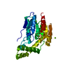 8g0nC  9c0mC  9c0nC C: citing same article ( |
|---|---|
| Similar structure data | |
| Other databases |
-Links
 PDBj
PDBj- Assembly
| Deposited unit | 
| ||||||||
|---|---|---|---|---|---|---|---|---|---|
| 1 |
| ||||||||
| Unit cell |
| ||||||||
| Components on special symmetry positions |
|
-Components
| #1: Protein | Mass: 28335.621 Da / Num. of mol.: 1 / Mutation: N-terminal GPG from expression tag Source method: isolated from a genetically manipulated source Source: (gene. exp.) Staphylococcus aureus USA300-CA-263 (bacteria)Gene: est_2 / Production host: | ||||||
|---|---|---|---|---|---|---|---|
| #2: Chemical | ChemComp-CA /   Mass: 40.078 Da / Num. of mol.: 4 / Source method: obtained synthetically / Formula: Ca Mass: 40.078 Da / Num. of mol.: 4 / Source method: obtained synthetically / Formula: Ca#3: Water | ChemComp-HOH / |  Mass: 18.015 Da / Num. of mol.: 145 / Source method: isolated from a natural source / Formula: H2O Mass: 18.015 Da / Num. of mol.: 145 / Source method: isolated from a natural source / Formula: H2OHas ligand of interest | N | Has protein modification | N | |
-Experimental details
-Experiment
| Experiment | Method: X-RAY DIFFRACTION / Number of used crystals: 1 |
|---|
- Sample preparation
| Crystal | Density Matthews: 2.69 Å3/Da / Density % sol: 54.3 % |
|---|---|
| Crystal grow | Temperature: 289.15 K / Method: vapor diffusion, sitting drop / pH: 8.5 Details: 0.3 uL 9.1 mg/mL FphH (10mM HEPES pH 7.6, 100mM NaCl) were mixed with 0.15 uL of reservoir solution. Sitting drop reservoir contained 200mM Calcium acetate hydrate, 100mM Tris pH 8.5, 25 % ...Details: 0.3 uL 9.1 mg/mL FphH (10mM HEPES pH 7.6, 100mM NaCl) were mixed with 0.15 uL of reservoir solution. Sitting drop reservoir contained 200mM Calcium acetate hydrate, 100mM Tris pH 8.5, 25 % w/v PEG 2000 MME. Crystal was frozen in a solution of ~25% Ethylenglycol, 75% reservoir. |
-Data collection
| Diffraction | Mean temperature: 100 K / Serial crystal experiment: N |
|---|---|
| Diffraction source | Source: SYNCHROTRON / Site: Australian Synchrotron  / Beamline: MX2 / Wavelength: 0.954 Å / Beamline: MX2 / Wavelength: 0.954 Å |
| Detector | Type: DECTRIS EIGER X 16M / Detector: PIXEL / Date: Apr 27, 2022 |
| Radiation | Protocol: SINGLE WAVELENGTH / Monochromatic (M) / Laue (L): M / Scattering type: x-ray |
| Radiation wavelength | Wavelength: 0.954 Å / Relative weight: 1 |
| Reflection | Resolution: 1.79→48.94 Å / Num. obs: 30069 / % possible obs: 99.7 % / Redundancy: 19.2 % / CC1/2: 0.999 / Rmerge(I) obs: 0.113 / Rpim(I) all: 0.027 / Rrim(I) all: 0.116 / Χ2: 1.01 / Net I/σ(I): 15.5 / Num. measured all: 576148 |
| Reflection shell | Resolution: 1.79→1.83 Å / % possible obs: 94.7 % / Redundancy: 18.6 % / Rmerge(I) obs: 2.565 / Num. measured all: 30217 / Num. unique obs: 1622 / CC1/2: 0.677 / Rpim(I) all: 0.593 / Rrim(I) all: 2.636 / Χ2: 0.94 / Net I/σ(I) obs: 1.4 |
- Processing
| Software |
| ||||||||||||||||||||||||||||||||||||||||||||||||||||||||||||||||||||||||||||||||||||||||||||||||||||
|---|---|---|---|---|---|---|---|---|---|---|---|---|---|---|---|---|---|---|---|---|---|---|---|---|---|---|---|---|---|---|---|---|---|---|---|---|---|---|---|---|---|---|---|---|---|---|---|---|---|---|---|---|---|---|---|---|---|---|---|---|---|---|---|---|---|---|---|---|---|---|---|---|---|---|---|---|---|---|---|---|---|---|---|---|---|---|---|---|---|---|---|---|---|---|---|---|---|---|---|---|---|
| Refinement | Method to determine structure: MOLECULAR REPLACEMENT / Resolution: 1.79→41.66 Å / SU ML: 0.21 / Cross valid method: FREE R-VALUE / σ(F): 1.34 / Phase error: 22.27 / Stereochemistry target values: ML
| ||||||||||||||||||||||||||||||||||||||||||||||||||||||||||||||||||||||||||||||||||||||||||||||||||||
| Solvent computation | Shrinkage radii: 0.9 Å / VDW probe radii: 1.1 Å / Solvent model: FLAT BULK SOLVENT MODEL | ||||||||||||||||||||||||||||||||||||||||||||||||||||||||||||||||||||||||||||||||||||||||||||||||||||
| Refinement step | Cycle: LAST / Resolution: 1.79→41.66 Å
| ||||||||||||||||||||||||||||||||||||||||||||||||||||||||||||||||||||||||||||||||||||||||||||||||||||
| Refine LS restraints |
| ||||||||||||||||||||||||||||||||||||||||||||||||||||||||||||||||||||||||||||||||||||||||||||||||||||
| LS refinement shell |
| ||||||||||||||||||||||||||||||||||||||||||||||||||||||||||||||||||||||||||||||||||||||||||||||||||||
| Refinement TLS params. | Method: refined / Refine-ID: X-RAY DIFFRACTION
| ||||||||||||||||||||||||||||||||||||||||||||||||||||||||||||||||||||||||||||||||||||||||||||||||||||
| Refinement TLS group |
|
