 Movie
Movie Controller

[English] 日本語
 Yorodumi
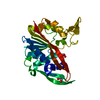
- PDB-9bxk: OvsM from Marinimicrobium koreense, an ovoselenol-biosynthetic N-... -
+ Open data

- Basic information
| Entry | Database: PDB / ID: 9bxk | |||||||||
|---|---|---|---|---|---|---|---|---|---|---|
| Title | OvsM from Marinimicrobium koreense, an ovoselenol-biosynthetic N-methyltransferase in complex with 5-selenohistidine and SAH | |||||||||
 Components Components | 5-selenohistidine N-methyltransferase OvsM | |||||||||
 Keywords Keywords | TRANSFERASE / ovoselenol / 5-selenohistidine / S-adenosyl-L-methionine / N-methyltransferase | |||||||||
| Function / homology | Putative 4-mercaptohistidine N1-methyltranferase, OvoA C-terminal domain / Methyltransferase domain / methyltransferase activity / methylation / S-adenosyl-L-methionine-dependent methyltransferase superfamily / : / S-ADENOSYL-L-HOMOCYSTEINE / Putative 4-mercaptohistidine N1-methyltransferase Function and homology information Function and homology information | |||||||||
| Biological species |  Marinimicrobium koreense (bacteria) Marinimicrobium koreense (bacteria) | |||||||||
| Method |  X-RAY DIFFRACTION / SYNCHROTRON / SIR / Resolution: 1.66 Å X-RAY DIFFRACTION / SYNCHROTRON / SIR / Resolution: 1.66 Å | |||||||||
 Authors Authors | Ireland, K.A. / Davis, K.M. | |||||||||
| Funding support |  United States, 2items United States, 2items
| |||||||||
 Citation Citation | Journal: Structure / Year: 2025 Title: Structural and functional analysis of SAM-dependent N-methyltransferases involved in ovoselenol and ovothiol biosynthesis. Authors: Ireland, K.A. / Kayrouz, C.M. / Abbott, M.L. / Seyedsayamdost, M.R. / Davis, K.M. | |||||||||
| History |
|
- Structure visualization
| Structure viewer | Molecule: MolmilJmol/JSmol |
|---|
- Downloads & links
-Download
| PDBx/mmCIF format | 9bxk.cif.gz | 78.5 KB | Display | PDBx/mmCIF format |
|---|---|---|---|---|
| PDB format | pdb9bxk.ent.gz | Display | PDB format | |
| PDBx/mmJSON format | 9bxk.json.gz | Tree view | PDBx/mmJSON format | |
| Others |  Other downloads Other downloads |
-Validation report
| Arichive directory | https://data.pdbj.org/pub/pdb/validation_reports/bx/9bxkftp://data.pdbj.org/pub/pdb/validation_reports/bx/9bxk | HTTPS FTP |
|---|
-Related structure data


-Links
 PDBj
PDBj- Assembly
| Deposited unit | 
| ||||||||||||
|---|---|---|---|---|---|---|---|---|---|---|---|---|---|
| 1 |
| ||||||||||||
| Unit cell |
| ||||||||||||
| Components on special symmetry positions |
|
-Components
| #1: Protein | Mass: 29997.824 Da / Num. of mol.: 1 Source method: isolated from a genetically manipulated source Source: (gene. exp.) Marinimicrobium koreense (bacteria) / Strain: DSM 16974 / Gene: EDC38_1804 / Production host: |
|---|---|
| #2: Chemical | ChemComp-SAH /   Mass: 384.411 Da / Num. of mol.: 1 / Source method: obtained synthetically / Formula: C14H20N6O5S / Feature type: SUBJECT OF INVESTIGATION Mass: 384.411 Da / Num. of mol.: 1 / Source method: obtained synthetically / Formula: C14H20N6O5S / Feature type: SUBJECT OF INVESTIGATION |
| #3: Chemical | ChemComp-A1AS0 / Type: L-peptide linking / Mass: 234.115 Da / Num. of mol.: 1 / Source method: obtained synthetically / Formula: C6H9N3O2Se / Feature type: SUBJECT OF INVESTIGATION |
| #4: Chemical | ChemComp-EDO /   Mass: 62.068 Da / Num. of mol.: 1 / Source method: obtained synthetically / Formula: C2H6O2 Mass: 62.068 Da / Num. of mol.: 1 / Source method: obtained synthetically / Formula: C2H6O2 |
| #5: Water | ChemComp-HOH /  Mass: 18.015 Da / Num. of mol.: 282 / Source method: isolated from a natural source / Formula: H2O Mass: 18.015 Da / Num. of mol.: 282 / Source method: isolated from a natural source / Formula: H2O |
| Has ligand of interest | Y |
| Has protein modification | N |
-Experimental details
-Experiment
| Experiment | Method: X-RAY DIFFRACTION / Number of used crystals: 1 |
|---|
- Sample preparation
| Crystal | Density Matthews: 2.5 Å3/Da / Density % sol: 50.8 % / Description: Plates |
|---|---|
| Crystal grow | Temperature: 293 K / Method: vapor diffusion, sitting drop / pH: 8.5 Details: 0.1 M Tris-HCl, pH 8.5, 0.2 M tri-sodium citrate, 30% v/v PEG400 |
-Data collection
| Diffraction | Mean temperature: 100 K / Serial crystal experiment: N |
|---|---|
| Diffraction source | Source: SYNCHROTRON / Site: CHESS / Beamline: 7B2 / Wavelength: 0.9686 Å |
| Detector | Type: DECTRIS EIGER2 S 16M / Detector: PIXEL / Date: Oct 19, 2023 |
| Radiation | Protocol: SINGLE WAVELENGTH / Monochromatic (M) / Laue (L): M / Scattering type: x-ray |
| Radiation wavelength | Wavelength: 0.9686 Å / Relative weight: 1 |
| Reflection | Resolution: 1.66→29 Å / Num. obs: 35794 / % possible obs: 98.75 % / Redundancy: 1.9 % / Biso Wilson estimate: 20.26 Å2 / CC1/2: 0.997 / CC star: 0.999 / Rmerge(I) obs: 0.05334 / Rpim(I) all: 0.05334 / Rrim(I) all: 0.07543 / Net I/σ(I): 9.95 |
| Reflection shell | Resolution: 1.66→1.719 Å / Redundancy: 1.9 % / Rmerge(I) obs: 0.6965 / Mean I/σ(I) obs: 1.2 / Num. unique obs: 3373 / CC1/2: 0.521 / CC star: 0.827 / Rpim(I) all: 0.6965 / Rrim(I) all: 0.985 / % possible all: 95.33 |
- Processing
| Software |
| |||||||||||||||||||||||||||||||||||||||||||||||||||||||||||||||||||||||||||||
|---|---|---|---|---|---|---|---|---|---|---|---|---|---|---|---|---|---|---|---|---|---|---|---|---|---|---|---|---|---|---|---|---|---|---|---|---|---|---|---|---|---|---|---|---|---|---|---|---|---|---|---|---|---|---|---|---|---|---|---|---|---|---|---|---|---|---|---|---|---|---|---|---|---|---|---|---|---|---|
| Refinement | Method to determine structure: SIR / Resolution: 1.66→29 Å / SU ML: 0.2028 / Cross valid method: FREE R-VALUE / σ(F): 1.34 / Phase error: 23.4549 Stereochemistry target values: GeoStd + Monomer Library + CDL v1.2
| |||||||||||||||||||||||||||||||||||||||||||||||||||||||||||||||||||||||||||||
| Solvent computation | Shrinkage radii: 0.9 Å / VDW probe radii: 1.1 Å / Solvent model: FLAT BULK SOLVENT MODEL | |||||||||||||||||||||||||||||||||||||||||||||||||||||||||||||||||||||||||||||
| Displacement parameters | Biso mean: 24.79 Å2 | |||||||||||||||||||||||||||||||||||||||||||||||||||||||||||||||||||||||||||||
| Refinement step | Cycle: LAST / Resolution: 1.66→29 Å
| |||||||||||||||||||||||||||||||||||||||||||||||||||||||||||||||||||||||||||||
| Refine LS restraints |
| |||||||||||||||||||||||||||||||||||||||||||||||||||||||||||||||||||||||||||||
| LS refinement shell |
|
