[English] 日本語
Yorodumi
- PDB-9box: Room-temperature X-ray structure of human mitochondrial serine hy... -

+
Open data


ID or keywords:

Loading...

-
Basic information

Entry
Database: PDB / ID: 9box
Title
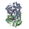
Room-temperature X-ray structure of human mitochondrial serine hydroxymethyltransferase (hSHMT2) with PLP-glycine external aldimine and 5-formyltetrahydrofolate (folinic acid)
ComponentsSerine hydroxymethyltransferase, mitochondrial
KeywordsTRANSFERASE / PYRIDOXAL 5'-PHOSPHATE / PLP / FOLD TYPE 1 / ONE CARBON METABOLISM
Function / homology
Function and homology information


BRISC complex / L-allo-threonine aldolase activity / regulation of mitochondrial translation / glycine metabolic process / L-serine metabolic process / regulation of oxidative phosphorylation / L-serine biosynthetic process / glycine hydroxymethyltransferase / glycine hydroxymethyltransferase activity / glycine biosynthetic process from serine ...BRISC complex / L-allo-threonine aldolase activity / regulation of mitochondrial translation / glycine metabolic process / L-serine metabolic process / regulation of oxidative phosphorylation / L-serine biosynthetic process / glycine hydroxymethyltransferase / glycine hydroxymethyltransferase activity / glycine biosynthetic process from serine / Metabolism of folate and pterines / tetrahydrofolate metabolic process / response to type I interferon / protein K63-linked deubiquitination / tetrahydrofolate interconversion / regulation of aerobic respiration / amino acid binding / mitochondrial nucleoid / RHOG GTPase cycle / Mitochondrial protein degradation / one-carbon metabolic process / protein tetramerization / pyridoxal phosphate binding / microtubule cytoskeleton / protein homotetramerization / mitochondrial inner membrane / mitochondrial matrix / positive regulation of cell population proliferation / chromatin binding / mitochondrion / extracellular exosome / identical protein binding / nucleus / cytoplasm
Similarity search - Function
Serine hydroxymethyltransferase, pyridoxal phosphate binding site / Serine hydroxymethyltransferase pyridoxal-phosphate attachment site. / : / Serine hydroxymethyltransferase / Serine hydroxymethyltransferase-like domain / Serine hydroxymethyltransferase / Pyridoxal phosphate-dependent transferase, small domain / Pyridoxal phosphate-dependent transferase, major domain / Pyridoxal phosphate-dependent transferase
Similarity search - Domain/homology
: / Chem-FFO / Serine hydroxymethyltransferase, mitochondrial
Similarity search - Component
Biological speciesHomo sapiens (human)
MethodX-RAY DIFFRACTION / SYNCHROTRON / MOLECULAR REPLACEMENT / Resolution: 2.1 Å
AuthorsDrago, V.N. / Kovalevsky, A.
Funding support United States, 1items
OrganizationGrant numberCountry
National Institutes of Health/National Institute of General Medical Sciences (NIH/NIGMS)R01GM137008 United States
CitationJournal: Chem Sci / Year: 2024
Title: Universality of critical active site glutamate as an acid-base catalyst in serine hydroxymethyltransferase function.
Authors: Drago, V.N. / Phillips, R.S. / Kovalevsky, A.
History
DepositionMay 6, 2024Deposition site: RCSB / Processing site: RCSB
Revision 1.0Aug 28, 2024Provider: repository / Type: Initial release

-
Structure visualization

Structure viewerMolecule:
MolmilJmol/JSmol

Downloads & links

-
Assembly

Deposited unit
A: Serine hydroxymethyltransferase, mitochondrial
B: Serine hydroxymethyltransferase, mitochondrial
C: Serine hydroxymethyltransferase, mitochondrial
D: Serine hydroxymethyltransferase, mitochondrial
hetero molecules


Theoretical massNumber of molelcules
Total (without water)208,42812
Polymers205,3184
Non-polymers3,1118
Water13,781765
1
A: Serine hydroxymethyltransferase, mitochondrial
C: Serine hydroxymethyltransferase, mitochondrial
hetero molecules


Theoretical massNumber of molelcules
Total (without water)104,2146
Polymers102,6592
Non-polymers1,5554
Water362
TypeNameSymmetry operationNumber
identity operation1_555x,y,z1
Buried area11520 Å2
ΔGint-49 kcal/mol
Surface area30870 Å2
MethodPISA
2
B: Serine hydroxymethyltransferase, mitochondrial
D: Serine hydroxymethyltransferase, mitochondrial
hetero molecules


Theoretical massNumber of molelcules
Total (without water)104,2146
Polymers102,6592
Non-polymers1,5554
Water362
TypeNameSymmetry operationNumber
identity operation1_555x,y,z1
Buried area11340 Å2
ΔGint-52 kcal/mol
Surface area30900 Å2
MethodPISA
Unit cell
Length a, b, c (Å)142.980, 119.300, 134.627
Angle α, β, γ (deg.)90.00, 90.00, 90.00
Int Tables number17
Space group name H-MP2221

-
Components

#1: Protein
Serine hydroxymethyltransferase, mitochondrial / SHMT / Glycine hydroxymethyltransferase / Serine methylase


Mass: 51329.453 Da / Num. of mol.: 4
Source method: isolated from a genetically manipulated source
Source: (gene. exp.) Homo sapiens (human) / Gene: SHMT2 / Production host: Escherichia coli (E. coli)
References: UniProt: P34897, glycine hydroxymethyltransferase
#2: Chemical
ChemComp-A1AQW / (E)-N-({3-hydroxy-2-methyl-5-[(phosphonooxy)methyl]pyridin-4-yl}methylidene)glycine


Mass: 304.193 Da / Num. of mol.: 4 / Source method: obtained synthetically / Formula: C10H13N2O7P / Feature type: SUBJECT OF INVESTIGATION
#3: Chemical
ChemComp-FFO / N-[4-({[(6S)-2-amino-5-formyl-4-oxo-3,4,5,6,7,8-hexahydropteridin-6-yl]methyl}amino)benzoyl]-L-glutamic acid / [6S]-5-FORMYL-TETRAHYDROFOLATE / 6S-FOLINIC ACID


Mass: 473.439 Da / Num. of mol.: 4 / Source method: obtained synthetically / Formula: C20H23N7O7 / Feature type: SUBJECT OF INVESTIGATION
#4: Water ChemComp-HOH / water


Mass: 18.015 Da / Num. of mol.: 765 / Source method: isolated from a natural source / Formula: H2O
Has ligand of interestY

-
Experimental details

-
Experiment

ExperimentMethod: X-RAY DIFFRACTION / Number of used crystals: 1

-
Sample preparation

CrystalDensity Matthews: 2.8 Å3/Da / Density % sol: 56.11 %
Crystal growTemperature: 293.15 K / Method: vapor diffusion, sitting drop / pH: 8.6 / Details: 0.5 M Gly pH 8.6, 10 mM folinic acid, 18% PEG 3350

-
Data collection

DiffractionMean temperature: 293 K / Serial crystal experiment: N
Diffraction sourceSource: SYNCHROTRON / Site: APS / Beamline: 19-ID / Wavelength: 0.979 Å
DetectorType: DECTRIS PILATUS 6M / Detector: PIXEL / Date: Feb 24, 2023
RadiationMonochromator: M / Protocol: SINGLE WAVELENGTH / Monochromatic (M) / Laue (L): M / Scattering type: x-ray
Radiation wavelengthWavelength: 0.979 Å / Relative weight: 1
ReflectionResolution: 2.1→50 Å / Num. obs: 125654 / % possible obs: 93.4 % / Redundancy: 4.4 % / CC1/2: 0.987 / Rmerge(I) obs: 0.113 / Rpim(I) all: 0.062 / Net I/σ(I): 12.9
Reflection shellResolution: 2.1→2.14 Å / Redundancy: 4.1 % / Rmerge(I) obs: 0.739 / Mean I/σ(I) obs: 2.2 / Num. unique obs: 6402 / CC1/2: 0.483 / Rpim(I) all: 0.427

-
Processing

Software
NameVersionClassification
PHENIX(1.20.1_4487: ???)refinement
HKL-3000data scaling
HKL-3000data reduction
PHASERphasing
RefinementMethod to determine structure: MOLECULAR REPLACEMENT / Resolution: 2.1→42.82 Å / SU ML: 0.21 / Cross valid method: THROUGHOUT / σ(F): 1 / Phase error: 20.44 / Stereochemistry target values: ML
RfactorNum. reflection% reflection
Rfree0.1886 6066 4.97 %
Rwork0.1623 --
obs0.1636 122017 90.64 %
Solvent computationShrinkage radii: 0.9 Å / VDW probe radii: 1.1 Å / Solvent model: FLAT BULK SOLVENT MODEL
Refinement stepCycle: LAST / Resolution: 2.1→42.82 Å
ProteinNucleic acidLigandSolventTotal
Num. atoms14292 0 216 765 15273
Refine LS restraints
Refine-IDTypeDev idealNumber
X-RAY DIFFRACTIONf_bond_d0.00914828
X-RAY DIFFRACTIONf_angle_d0.97320066
X-RAY DIFFRACTIONf_dihedral_angle_d16.2725572
X-RAY DIFFRACTIONf_chiral_restr0.0512184
X-RAY DIFFRACTIONf_plane_restr0.0142628
LS refinement shell
Resolution (Å)Rfactor RfreeNum. reflection RfreeRfactor RworkNum. reflection RworkRefine-ID% reflection obs (%)
2.1-2.120.27491710.25093412X-RAY DIFFRACTION80
2.12-2.150.23632030.23163532X-RAY DIFFRACTION84
2.15-2.170.25161680.23373605X-RAY DIFFRACTION85
2.17-2.20.25581810.22873676X-RAY DIFFRACTION87
2.2-2.230.25341990.23233717X-RAY DIFFRACTION88
2.23-2.260.27362090.22113774X-RAY DIFFRACTION89
2.26-2.290.26342110.21543854X-RAY DIFFRACTION91
2.29-2.330.22161920.21163879X-RAY DIFFRACTION92
2.33-2.360.24571880.21273928X-RAY DIFFRACTION92
2.36-2.40.26471940.21163892X-RAY DIFFRACTION92
2.4-2.440.23381970.20533946X-RAY DIFFRACTION92
2.44-2.490.22812140.20323927X-RAY DIFFRACTION93
2.49-2.540.24152030.20223954X-RAY DIFFRACTION93
2.54-2.590.21452020.20053946X-RAY DIFFRACTION93
2.59-2.640.22522210.19463948X-RAY DIFFRACTION93
2.65-2.710.24822250.18663937X-RAY DIFFRACTION93
2.71-2.770.22272240.17953948X-RAY DIFFRACTION93
2.77-2.850.22122240.17723924X-RAY DIFFRACTION93
2.85-2.930.19852230.17123945X-RAY DIFFRACTION93
2.93-3.030.19531680.16164002X-RAY DIFFRACTION93
3.03-3.140.19972150.16283942X-RAY DIFFRACTION93
3.14-3.260.18411960.15273969X-RAY DIFFRACTION92
3.26-3.410.17671970.14553957X-RAY DIFFRACTION92
3.41-3.590.15722080.13283948X-RAY DIFFRACTION92
3.59-3.810.14681970.12223901X-RAY DIFFRACTION91
3.81-4.110.12581920.11553944X-RAY DIFFRACTION91
4.11-4.520.13122060.11253882X-RAY DIFFRACTION90
4.52-5.170.13532190.11563894X-RAY DIFFRACTION89
5.18-6.520.16112090.14173860X-RAY DIFFRACTION89
6.52-42.820.14262100.13093908X-RAY DIFFRACTION86

+
About Yorodumi

-
News

-
Feb 9, 2022. New format data for meta-information of EMDB entries

New format data for meta-information of EMDB entries

  • Version 3 of the EMDB header file is now the official format.
  • The previous official version 1.9 will be removed from the archive.

Related info.:EMDB header

External links:wwPDB to switch to version 3 of the EMDB data model

-
Aug 12, 2020. Covid-19 info

Covid-19 info

URL: https://pdbj.org/emnavi/covid19.php

New page: Covid-19 featured information page in EM Navigator.

Related info.:Covid-19 info / Mar 5, 2020. Novel coronavirus structure data

+
Mar 5, 2020. Novel coronavirus structure data

Novel coronavirus structure data

Related info.:Yorodumi Speices / Aug 12, 2020. Covid-19 info

External links:COVID-19 featured content - PDBj / Molecule of the Month (242):Coronavirus Proteases

+
Jan 31, 2019. EMDB accession codes are about to change! (news from PDBe EMDB page)

EMDB accession codes are about to change! (news from PDBe EMDB page)

  • The allocation of 4 digits for EMDB accession codes will soon come to an end. Whilst these codes will remain in use, new EMDB accession codes will include an additional digit and will expand incrementally as the available range of codes is exhausted. The current 4-digit format prefixed with “EMD-” (i.e. EMD-XXXX) will advance to a 5-digit format (i.e. EMD-XXXXX), and so on. It is currently estimated that the 4-digit codes will be depleted around Spring 2019, at which point the 5-digit format will come into force.
  • The EM Navigator/Yorodumi systems omit the EMD- prefix.

Related info.:Q: What is EMD? / ID/Accession-code notation in Yorodumi/EM Navigator

External links:EMDB Accession Codes are Changing Soon! / Contact to PDBj

+
Jul 12, 2017. Major update of PDB

Major update of PDB

  • wwPDB released updated PDB data conforming to the new PDBx/mmCIF dictionary.
  • This is a major update changing the version number from 4 to 5, and with Remediation, in which all the entries are updated.
  • In this update, many items about electron microscopy experimental information are reorganized (e.g. em_software).
  • Now, EM Navigator and Yorodumi are based on the updated data.

External links:wwPDB Remediation / Enriched Model Files Conforming to OneDep Data Standards Now Available in the PDB FTP Archive

-
Yorodumi

Thousand views of thousand structures

  • Yorodumi is a browser for structure data from EMDB, PDB, SASBDB, etc.
  • This page is also the successor to EM Navigator detail page, and also detail information page/front-end page for Omokage search.
  • The word "yorodu" (or yorozu) is an old Japanese word meaning "ten thousand". "mi" (miru) is to see.

Related info.:EMDB / PDB / SASBDB / Comparison of 3 databanks / Yorodumi Search / Aug 31, 2016. New EM Navigator & Yorodumi / Yorodumi Papers / Jmol/JSmol / Function and homology information / Changes in new EM Navigator and Yorodumi

Read more