 Movie
Movie Controller

[English] 日本語
 Yorodumi
- PDB-9bow: X-ray structure of Thermus thermophilus serine hydroxymethyltrans... -
+ Open data

- Basic information
| Entry | Database: PDB / ID: 9bow | ||||||
|---|---|---|---|---|---|---|---|
| Title | X-ray structure of Thermus thermophilus serine hydroxymethyltransferase with PLP-L-Ser external aldimine and 5-formyltetrahydrofolate (folinic acid) | ||||||
 Components Components | (Serine hydroxymethyltransferase) x 2 | ||||||
 Keywords Keywords | TRANSFERASE / PYRIDOXAL 5'-PHOSPHATE / PLP / FOLD TYPE 1 / ONE CARBON METABOLISM | ||||||
| Function / homology |  Function and homology information Function and homology informationglycine hydroxymethyltransferase / glycine hydroxymethyltransferase activity / : / tetrahydrofolate interconversion / pyridoxal phosphate binding / cytosol Similarity search - Function | ||||||
| Biological species |   Thermus thermophilus HB8 (bacteria) Thermus thermophilus HB8 (bacteria) | ||||||
| Method |  X-RAY DIFFRACTION / MOLECULAR REPLACEMENT / Resolution: 1.8 Å X-RAY DIFFRACTION / MOLECULAR REPLACEMENT / Resolution: 1.8 Å | ||||||
 Authors Authors | Drago, V.N. / Kovalevsky, A. | ||||||
| Funding support |  United States, 1items United States, 1items
| ||||||
 Citation Citation | Journal: Chem Sci / Year: 2024 Title: Universality of critical active site glutamate as an acid-base catalyst in serine hydroxymethyltransferase function. Authors: Drago, V.N. / Phillips, R.S. / Kovalevsky, A. | ||||||
| History |
|
- Structure visualization
| Structure viewer | Molecule: MolmilJmol/JSmol |
|---|
- Downloads & links
-Download
| PDBx/mmCIF format | 9bow.cif.gz | 191.2 KB | Display | PDBx/mmCIF format |
|---|---|---|---|---|
| PDB format | pdb9bow.ent.gz | 145.7 KB | Display | PDB format |
| PDBx/mmJSON format | 9bow.json.gz | Tree view | PDBx/mmJSON format | |
| Others |  Other downloads Other downloads |
-Validation report
| Arichive directory | https://data.pdbj.org/pub/pdb/validation_reports/bo/9bowftp://data.pdbj.org/pub/pdb/validation_reports/bo/9bow | HTTPS FTP |
|---|
-Related structure data


-Links
 PDBj
PDBj- Assembly
| Deposited unit | 
| ||||||||
|---|---|---|---|---|---|---|---|---|---|
| 1 |
| ||||||||
| Unit cell |
|
-Components
-Protein , 2 types, 2 molecules AB
| #1: Protein | Mass: 44376.684 Da / Num. of mol.: 1 Source method: isolated from a genetically manipulated source Source: (gene. exp.) Thermus thermophilus HB8 (bacteria) / Gene: glyA, TTHA1524 / Production host: References: UniProt: Q5SI56, glycine hydroxymethyltransferase |
|---|---|
| #2: Protein | Mass: 44148.562 Da / Num. of mol.: 1 Source method: isolated from a genetically manipulated source Source: (gene. exp.) Thermus thermophilus HB8 (bacteria) / Gene: glyA, TTHA1524 / Production host: References: UniProt: Q5SI56, glycine hydroxymethyltransferase |
-Non-polymers , 5 types, 831 molecules 

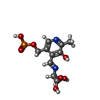


| #3: Chemical | ChemComp-SER /  Type: L-peptide linking / Mass: 105.093 Da / Num. of mol.: 1 / Source method: obtained synthetically / Formula: C3H7NO3 / Feature type: SUBJECT OF INVESTIGATION Type: L-peptide linking / Mass: 105.093 Da / Num. of mol.: 1 / Source method: obtained synthetically / Formula: C3H7NO3 / Feature type: SUBJECT OF INVESTIGATION | ||||||
|---|---|---|---|---|---|---|---|
| #4: Chemical |  Mass: 96.063 Da / Num. of mol.: 2 / Source method: obtained synthetically / Formula: SO4 Mass: 96.063 Da / Num. of mol.: 2 / Source method: obtained synthetically / Formula: SO4#5: Chemical | ChemComp-KOU / ( |  Mass: 334.219 Da / Num. of mol.: 1 / Source method: obtained synthetically / Formula: C11H15N2O8P / Feature type: SUBJECT OF INVESTIGATION Mass: 334.219 Da / Num. of mol.: 1 / Source method: obtained synthetically / Formula: C11H15N2O8P / Feature type: SUBJECT OF INVESTIGATION#6: Chemical | ChemComp-FFO / |  Mass: 473.439 Da / Num. of mol.: 1 / Source method: obtained synthetically / Formula: C20H23N7O7 / Feature type: SUBJECT OF INVESTIGATION Mass: 473.439 Da / Num. of mol.: 1 / Source method: obtained synthetically / Formula: C20H23N7O7 / Feature type: SUBJECT OF INVESTIGATION#7: Water | ChemComp-HOH / | Mass: 18.015 Da / Num. of mol.: 826 / Source method: isolated from a natural source / Formula: H2O |
-Details
| Has ligand of interest | Y |
|---|
-Experimental details
-Experiment
| Experiment | Method: X-RAY DIFFRACTION / Number of used crystals: 1 |
|---|
- Sample preparation
| Crystal | Density Matthews: 2.57 Å3/Da / Density % sol: 52.08 % |
|---|---|
| Crystal grow | Temperature: 289.15 K / Method: vapor diffusion, sitting drop / pH: 5.5 Details: 40 mM NaOAc pH 5.5, 1 M (NH4)2SO4, 0.5 M Li2SO4, soaked in 40 mM NaOAc pH 5.5, 0.5 M L-Ser, 15% PEG 4000 |
-Data collection
| Diffraction | Mean temperature: 293 K / Serial crystal experiment: N |
|---|---|
| Diffraction source | Source: ROTATING ANODE / Type: RIGAKU MICROMAX-007 HF / Wavelength: 1.5406 Å |
| Detector | Type: DECTRIS EIGER R 4M / Detector: PIXEL / Date: Jan 10, 2023 / Details: Osmic VariMax |
| Radiation | Monochromator: M / Protocol: SINGLE WAVELENGTH / Monochromatic (M) / Laue (L): M / Scattering type: x-ray |
| Radiation wavelength | Wavelength: 1.5406 Å / Relative weight: 1 |
| Reflection | Resolution: 1.8→29.05 Å / Num. obs: 76390 / % possible obs: 92.2 % / Redundancy: 3.5 % / CC1/2: 0.997 / Rmerge(I) obs: 0.03 / Rpim(I) all: 0.026 / Net I/σ(I): 23.3 |
| Reflection shell | Resolution: 1.8→1.84 Å / Redundancy: 2.2 % / Rmerge(I) obs: 0.111 / Mean I/σ(I) obs: 7.6 / Num. unique obs: 2744 / CC1/2: 0.969 / Rpim(I) all: 0.105 / % possible all: 56.4 |
- Processing
| Software |
| ||||||||||||||||||||||||||||||||||||||||||||||||||||||||||||||||||||||||||||||||||||||||||||||||||||||||||||||||||||||||||||||||||||||||||||||||||||||||||||||||||||||||||||||||||||||||||||||||||||
|---|---|---|---|---|---|---|---|---|---|---|---|---|---|---|---|---|---|---|---|---|---|---|---|---|---|---|---|---|---|---|---|---|---|---|---|---|---|---|---|---|---|---|---|---|---|---|---|---|---|---|---|---|---|---|---|---|---|---|---|---|---|---|---|---|---|---|---|---|---|---|---|---|---|---|---|---|---|---|---|---|---|---|---|---|---|---|---|---|---|---|---|---|---|---|---|---|---|---|---|---|---|---|---|---|---|---|---|---|---|---|---|---|---|---|---|---|---|---|---|---|---|---|---|---|---|---|---|---|---|---|---|---|---|---|---|---|---|---|---|---|---|---|---|---|---|---|---|---|---|---|---|---|---|---|---|---|---|---|---|---|---|---|---|---|---|---|---|---|---|---|---|---|---|---|---|---|---|---|---|---|---|---|---|---|---|---|---|---|---|---|---|---|---|---|---|---|---|
| Refinement | Method to determine structure: MOLECULAR REPLACEMENT / Resolution: 1.8→29.05 Å / SU ML: 0.12 / Cross valid method: THROUGHOUT / σ(F): 1.07 / Phase error: 15.15 / Stereochemistry target values: ML
| ||||||||||||||||||||||||||||||||||||||||||||||||||||||||||||||||||||||||||||||||||||||||||||||||||||||||||||||||||||||||||||||||||||||||||||||||||||||||||||||||||||||||||||||||||||||||||||||||||||
| Solvent computation | Shrinkage radii: 0.9 Å / VDW probe radii: 1.1 Å / Solvent model: FLAT BULK SOLVENT MODEL | ||||||||||||||||||||||||||||||||||||||||||||||||||||||||||||||||||||||||||||||||||||||||||||||||||||||||||||||||||||||||||||||||||||||||||||||||||||||||||||||||||||||||||||||||||||||||||||||||||||
| Refinement step | Cycle: LAST / Resolution: 1.8→29.05 Å
| ||||||||||||||||||||||||||||||||||||||||||||||||||||||||||||||||||||||||||||||||||||||||||||||||||||||||||||||||||||||||||||||||||||||||||||||||||||||||||||||||||||||||||||||||||||||||||||||||||||
| Refine LS restraints |
| ||||||||||||||||||||||||||||||||||||||||||||||||||||||||||||||||||||||||||||||||||||||||||||||||||||||||||||||||||||||||||||||||||||||||||||||||||||||||||||||||||||||||||||||||||||||||||||||||||||
| LS refinement shell |
|
