[English] 日本語
 Yorodumi
- PDB-9be8: Alkalihalobacillus halodurans (Aha) trp RNA binding attenuation p... -
+ Open data
- Basic information
| Entry | Database: PDB / ID: 9be8 | |||||||||||||||||||||
|---|---|---|---|---|---|---|---|---|---|---|---|---|---|---|---|---|---|---|---|---|---|---|
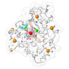
| Title | Alkalihalobacillus halodurans (Aha) trp RNA binding attenuation protein (TRAP) mutant T49A/T52A dTRAP with Trp | |||||||||||||||||||||
 Components Components | Transcription attenuation protein MtrB | |||||||||||||||||||||
 Keywords Keywords | RNA BINDING PROTEIN / Hexamer of T49A/T52A dTRAP with Trp | |||||||||||||||||||||
| Function / homology | Transcription attenuation protein MtrB / Tryptophan RNA-binding attenuator protein domain / Tryptophan RNA-binding attenuator protein / Tryptophan RNA-binding attenuator protein-like domain superfamily / DNA-templated transcription termination / regulation of DNA-templated transcription / RNA binding / TRYPTOPHAN / Transcription attenuation protein MtrB Function and homology information Function and homology information | |||||||||||||||||||||
| Biological species |  Halalkalibacterium halodurans (bacteria) Halalkalibacterium halodurans (bacteria) | |||||||||||||||||||||
| Method | ELECTRON MICROSCOPY / single particle reconstruction / cryo EM / Resolution: 4.14 Å | |||||||||||||||||||||
 Authors Authors | Yang, H. / Stachowski, K. / Foster, M. | |||||||||||||||||||||
| Funding support |  United States, 4items United States, 4items
| |||||||||||||||||||||
 Citation Citation |  Journal: Proc Natl Acad Sci U S A / Year: 2025 Journal: Proc Natl Acad Sci U S A / Year: 2025Title: Structural basis of nearest-neighbor cooperativity in the ring-shaped gene regulatory protein TRAP from protein engineering and cryo-EM. Authors: Weicheng Li / Haoyun Yang / Kye Stachowski / Andrew S Norris / Katie Lichtenthal / Skyler Kelly / Paul Gollnick / Vicki H Wysocki / Mark P Foster /  Abstract: The homo-dodecameric ring-shaped RNA binding attenuation protein (TRAP) from binds up to twelve tryptophan ligands (Trp) and becomes activated to bind a specific sequence in the 5' leader region of ...The homo-dodecameric ring-shaped RNA binding attenuation protein (TRAP) from binds up to twelve tryptophan ligands (Trp) and becomes activated to bind a specific sequence in the 5' leader region of the operon mRNA, thereby downregulating biosynthesis of Trp. Thermodynamic measurements of Trp binding have revealed a range of cooperative behavior for different TRAP variants, even if the averaged apparent affinities for Trp have been found to be similar. Proximity between the ligand binding sites, and the ligand-coupled disorder-to-order transition has implicated nearest-neighbor interactions in cooperativity. To establish a solid basis for describing nearest-neighbor cooperativity in TRAP, we engineered variants constructed with two subunits connected by a flexible linker (dTRAP). We mutated the binding sites of alternating protomers such that only every other site was competent for Trp binding (WT-Mut dTRAP). Ligand binding monitored by NMR, calorimetry, and native mass spectrometry revealed strong cooperativity in dTRAP containing adjacent binding-competent sites, but a severe binding defect when the wild-type sites were separated by mutated sites. Cryo-EM experiments of dTRAP in its ligand-free apo state, and both dTRAP and WT-Mut dTRAP in the presence of Trp, revealed progressive stabilization of loops that gate the Trp binding site and participate in RNA binding. These studies provide important insights into the thermodynamic and structural basis for the observed ligand binding cooperativity in TRAP. Such insights can be useful for understanding allosteric control networks and for the development of those with defined ligand sensitivity and regulatory control. | |||||||||||||||||||||
| History |
|
- Structure visualization
| Structure viewer | Molecule:  Molmil Molmil Jmol/JSmol Jmol/JSmol |
|---|
- Downloads & links
- Download
| PDBx/mmCIF format |  9be8.cif.gz 9be8.cif.gz | 273.1 KB | Display |  PDBx/mmCIF format PDBx/mmCIF format |
|---|---|---|---|---|
| PDB format |  pdb9be8.ent.gz pdb9be8.ent.gz | 222.3 KB | Display |  PDB format PDB format |
| PDBx/mmJSON format |  9be8.json.gz 9be8.json.gz | Tree view |  PDBx/mmJSON format PDBx/mmJSON format | |
| Others |  Other downloads Other downloads |
-Validation report
| Arichive directory |  https://data.pdbj.org/pub/pdb/validation_reports/be/9be8 https://data.pdbj.org/pub/pdb/validation_reports/be/9be8 ftp://data.pdbj.org/pub/pdb/validation_reports/be/9be8 ftp://data.pdbj.org/pub/pdb/validation_reports/be/9be8 | HTTPS FTP |
|---|
-Related structure data
| Related structure data |  44473MC  9bdsC  9be7C M: map data used to model this data C: citing same article ( |
|---|---|
| Similar structure data | Similarity search - Function & homology  F&H Search F&H Search |
- Links
Links
- Assembly
| Deposited unit | 
|
|---|---|
| 1 |
|
- Components
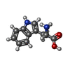
| #1: Protein | Mass: 18358.420 Da / Num. of mol.: 6 / Mutation: T49A,T52A Source method: isolated from a genetically manipulated source Source: (gene. exp.)  Halalkalibacterium halodurans (bacteria) Halalkalibacterium halodurans (bacteria)Gene: mtrB, BH1647 / Production host:  #2: Chemical | ChemComp-TRP / Has ligand of interest | Y | Has protein modification | N | |
|---|
-Experimental details
-Experiment
| Experiment | Method: ELECTRON MICROSCOPY |
|---|---|
| EM experiment | Aggregation state: PARTICLE / 3D reconstruction method: single particle reconstruction |
- Sample preparation
| Component | Name: Hexamer dTRAP T49A/T52A protein complex with Trp / Type: COMPLEX / Entity ID: #1 / Source: RECOMBINANT | |||||||||||||||
|---|---|---|---|---|---|---|---|---|---|---|---|---|---|---|---|---|
| Molecular weight | Experimental value: NO | |||||||||||||||
| Source (natural) | Organism:  Halalkalibacterium halodurans (bacteria) Halalkalibacterium halodurans (bacteria) | |||||||||||||||
| Source (recombinant) | Organism:  | |||||||||||||||
| Buffer solution | pH: 8 / Details: 20 mM HEPES, 200 mM NaCl, pH 8 | |||||||||||||||
| Buffer component |
| |||||||||||||||
| Specimen | Conc.: 6 mg/ml / Embedding applied: NO / Shadowing applied: NO / Staining applied: NO / Vitrification applied: YES Details: The sample of Trp-bound WT-Mut dTRAP was prepared by diluting 14 mg/mL of WT-Mut dTRAP with 20 mM Trp in cryo-EM buffer and 0.6% Triton X-100 to obtain a final protein concentration of 6 ...Details: The sample of Trp-bound WT-Mut dTRAP was prepared by diluting 14 mg/mL of WT-Mut dTRAP with 20 mM Trp in cryo-EM buffer and 0.6% Triton X-100 to obtain a final protein concentration of 6 mg/mL with 0.05% Triton X-100 and 1.4 mM Trp. | |||||||||||||||
| Specimen support | Details: 20 mA, 30 sec hold, 1 min glow discharge using a PELCO easiGlow Discharge System Grid material: GOLD / Grid mesh size: 300 divisions/in. / Grid type: Quantifoil R1.2/1.3 | |||||||||||||||
| Vitrification | Instrument: FEI VITROBOT MARK IV / Cryogen name: ETHANE / Humidity: 100 % / Chamber temperature: 277.15 K Details: lotted with a blot force of 1 for 4 seconds at 277.15K and 100% relative humidity before being plunged frozen into liquid ethane |
- Electron microscopy imaging
| Experimental equipment |  Model: Titan Krios / Image courtesy: FEI Company |
|---|---|
| Microscopy | Model: TFS KRIOS |
| Electron gun | Electron source:  FIELD EMISSION GUN / Accelerating voltage: 300 kV / Illumination mode: OTHER FIELD EMISSION GUN / Accelerating voltage: 300 kV / Illumination mode: OTHER |
| Electron lens | Mode: BRIGHT FIELD / Nominal defocus max: 2400 nm / Nominal defocus min: 1000 nm |
| Image recording | Electron dose: 60 e/Å2 / Film or detector model: GATAN K3 BIOCONTINUUM (6k x 4k) |
- Processing
| EM software | Name: PHENIX / Category: model refinement | ||||||||||||||||||||||||
|---|---|---|---|---|---|---|---|---|---|---|---|---|---|---|---|---|---|---|---|---|---|---|---|---|---|
| CTF correction | Type: PHASE FLIPPING AND AMPLITUDE CORRECTION | ||||||||||||||||||||||||
| 3D reconstruction | Resolution: 4.14 Å / Resolution method: FSC 0.143 CUT-OFF / Num. of particles: 48696 / Symmetry type: POINT | ||||||||||||||||||||||||
| Refine LS restraints |
|
 Movie
Movie Controller


 PDBj
PDBj


