Movie
Movie Controller

[English] 日本語
 Yorodumi
- PDB-9bbn: Cryo-EM structure of Chikungunya virus asymmetric unit with Fab C9 -
+ Open data

- Basic information
| Entry | Database: PDB / ID: 9bbn | ||||||||||||||||||||||||||||||
|---|---|---|---|---|---|---|---|---|---|---|---|---|---|---|---|---|---|---|---|---|---|---|---|---|---|---|---|---|---|---|---|
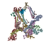
| Title | Cryo-EM structure of Chikungunya virus asymmetric unit with Fab C9 | ||||||||||||||||||||||||||||||
 Components Components |
| ||||||||||||||||||||||||||||||
 Keywords Keywords | VIRAL PROTEIN/IMMUNE SYSTEM / CHIKV asymmetric unit with Fab C9 / VIRAL PROTEIN-IMMUNE SYSTEM complex | ||||||||||||||||||||||||||||||
| Function / homology |  Function and homology information Function and homology informationT=4 icosahedral viral capsid / host cell endoplasmic reticulum / channel activity / monoatomic ion transmembrane transport / host cell Golgi apparatus / entry receptor-mediated virion attachment to host cell / serine-type endopeptidase activity / fusion of virus membrane with host endosome membrane / symbiont entry into host cell / host cell nucleus ...T=4 icosahedral viral capsid / host cell endoplasmic reticulum / channel activity / monoatomic ion transmembrane transport / host cell Golgi apparatus / entry receptor-mediated virion attachment to host cell / serine-type endopeptidase activity / fusion of virus membrane with host endosome membrane / symbiont entry into host cell / host cell nucleus / host cell plasma membrane / virion membrane / structural molecule activity / proteolysis / RNA binding Similarity search - Function | ||||||||||||||||||||||||||||||
| Biological species |    Chikungunya virus Chikungunya virus | ||||||||||||||||||||||||||||||
| Method | ELECTRON MICROSCOPY / single particle reconstruction / cryo EM / Resolution: 3.3 Å | ||||||||||||||||||||||||||||||
 Authors Authors | Su, G.C. / Galaz-Montoya, J.G. / Pintilie, G. / Jin, J. / Chiu, W. | ||||||||||||||||||||||||||||||
| Funding support |  United States, 1items United States, 1items
| ||||||||||||||||||||||||||||||
 Citation Citation | Journal: To Be Published Title: Cryogenic Electron Microscopy Reveals Dual-Inhibition Mechanisms and Explains the Differential Neutralization Potency of Two anti-CHIKV Antibodies Authors: Su, G.C. / Galaz-Montoya, J.G. / Pintilie, G. / Jin, J. / Chiu, W. | ||||||||||||||||||||||||||||||
| History |
|
- Structure visualization
| Structure viewer | Molecule: MolmilJmol/JSmol |
|---|
- Downloads & links
-Download
| PDBx/mmCIF format | 9bbn.cif.gz | 1.8 MB | Display | PDBx/mmCIF format |
|---|---|---|---|---|
| PDB format | pdb9bbn.ent.gz | 1.5 MB | Display | PDB format |
| PDBx/mmJSON format | 9bbn.json.gz | Tree view | PDBx/mmJSON format | |
| Others |  Other downloads Other downloads |
-Validation report
| Arichive directory | https://data.pdbj.org/pub/pdb/validation_reports/bb/9bbnftp://data.pdbj.org/pub/pdb/validation_reports/bb/9bbn | HTTPS FTP |
|---|
-Related structure data
| Related structure data |  44423MC  9bhhC M: map data used to model this data C: citing same article ( |
|---|---|
| Similar structure data |


-Links
 PDBj
PDBj


- Assembly
| Deposited unit | 
|
|---|---|
| 1 |
|
-Components
-Protein , 4 types, 16 molecules UWXVCABDGEFHJKIL
| #1: Protein | Mass: 6955.070 Da / Num. of mol.: 4 / Fragment: UNP residues 266-325 / Source method: isolated from a natural source / Source: (natural) Chikungunya virus / Strain: vaccine strain 181/clone / References: UniProt: D2KBQ2, togavirin#2: Protein | Mass: 47497.906 Da / Num. of mol.: 4 / Fragment: UNP residues 810-1248 / Source method: isolated from a natural source / Source: (natural) Chikungunya virus / Strain: vaccine strain 181/clone / References: UniProt: Q88628, togavirin#3: Protein | Mass: 47077.719 Da / Num. of mol.: 4 / Fragment: UNP residues 330-748 / Source method: isolated from a natural source / Source: (natural) Chikungunya virus / Strain: vaccine strain 181/clone 25 / References: UniProt: Q88628, togavirin#6: Protein | Mass: 16428.607 Da / Num. of mol.: 4 / Fragment: UNP residues 111-261 / Source method: isolated from a natural source / Source: (natural) Chikungunya virus / Strain: vaccine strain 181/clone 25 / References: UniProt: Q88628, togavirin |
|---|
-Antibody , 2 types, 8 molecules MOQSNPRT
| #4: Antibody | Mass: 24747.697 Da / Num. of mol.: 4 Source method: isolated from a genetically manipulated source Source: (gene. exp.)  Homo sapiens (human) Homo sapiens (human)#5: Antibody | Mass: 23061.602 Da / Num. of mol.: 4 Source method: isolated from a genetically manipulated source Source: (gene. exp.) Homo sapiens (human) |
|---|
-Details
| Has protein modification | Y |
|---|
-Experimental details
-Experiment
| Experiment | Method: ELECTRON MICROSCOPY |
|---|---|
| EM experiment | Aggregation state: PARTICLE / 3D reconstruction method: single particle reconstruction |
- Sample preparation
| Component | Name: Chikungunya virus / Type: VIRUS / Entity ID: all / Source: NATURAL | ||||||||||||||||
|---|---|---|---|---|---|---|---|---|---|---|---|---|---|---|---|---|---|
| Molecular weight | Value: 0.5 MDa / Experimental value: NO | ||||||||||||||||
| Source (natural) | Organism: Chikungunya virus / Strain: vaccine strain 181/clone 25 | ||||||||||||||||
| Details of virus | Empty: NO / Enveloped: YES / Isolate: STRAIN / Type: VIRION | ||||||||||||||||
| Virus shell | Diameter: 700 nm / Triangulation number (T number): 4 | ||||||||||||||||
| Buffer solution | pH: 8 | ||||||||||||||||
| Buffer component |
| ||||||||||||||||
| Specimen | Embedding applied: NO / Shadowing applied: NO / Staining applied: NO / Vitrification applied: YES | ||||||||||||||||
| Specimen support | Grid material: COPPER / Grid mesh size: 200 divisions/in. / Grid type: Quantifoil R2/1 | ||||||||||||||||
| Vitrification | Instrument: LEICA EM GP / Cryogen name: ETHANE / Humidity: 100 % / Chamber temperature: 293.15 K |
- Electron microscopy imaging
| Experimental equipment |  Model: Titan Krios / Image courtesy: FEI Company |
|---|---|
| Microscopy | Model: TFS KRIOS |
| Electron gun | Electron source:  FIELD EMISSION GUN / Accelerating voltage: 300 kV / Illumination mode: FLOOD BEAM FIELD EMISSION GUN / Accelerating voltage: 300 kV / Illumination mode: FLOOD BEAM |
| Electron lens | Mode: BRIGHT FIELD / Nominal magnification: 106000 X / Nominal defocus max: 2400 nm / Nominal defocus min: 1000 nm / Cs: 2.7 mm / C2 aperture diameter: 70 µm / Alignment procedure: COMA FREE |
| Specimen holder | Cryogen: NITROGEN / Specimen holder model: FEI TITAN KRIOS AUTOGRID HOLDER |
| Image recording | Electron dose: 45 e/Å2 / Detector mode: COUNTING / Film or detector model: GATAN K2 SUMMIT (4k x 4k) |
| EM imaging optics | Energyfilter name: GIF Bioquantum / Energyfilter slit width: 20 eV |
| Image scans | Movie frames/image: 40 |
- Processing
| EM software |
| ||||||||||||||||||||||||||||||||||||||||||||
|---|---|---|---|---|---|---|---|---|---|---|---|---|---|---|---|---|---|---|---|---|---|---|---|---|---|---|---|---|---|---|---|---|---|---|---|---|---|---|---|---|---|---|---|---|---|
| CTF correction | Type: PHASE FLIPPING AND AMPLITUDE CORRECTION | ||||||||||||||||||||||||||||||||||||||||||||
| Particle selection | Num. of particles selected: 192895 | ||||||||||||||||||||||||||||||||||||||||||||
| Symmetry | Point symmetry: C1 (asymmetric) | ||||||||||||||||||||||||||||||||||||||||||||
| 3D reconstruction | Resolution: 3.3 Å / Resolution method: FSC 0.143 CUT-OFF / Num. of particles: 1151475 / Num. of class averages: 1 / Symmetry type: POINT | ||||||||||||||||||||||||||||||||||||||||||||
| Atomic model building | Protocol: FLEXIBLE FIT / Space: REAL | ||||||||||||||||||||||||||||||||||||||||||||
| Atomic model building | PDB-ID: 8FCG Accession code: 8FCG / Source name: PDB / Type: experimental model | ||||||||||||||||||||||||||||||||||||||||||||
| Refinement | Highest resolution: 3.3 Å Stereochemistry target values: REAL-SPACE (WEIGHTED MAP SUM AT ATOM CENTERS) | ||||||||||||||||||||||||||||||||||||||||||||
| Refine LS restraints |
|