 Movie
Movie Controller


+ Open data

- Basic information
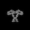
| Entry | Database: PDB / ID: 9ba5 | ||||||
|---|---|---|---|---|---|---|---|
| Title | Cross-linked Contactin 2 Ig1-Ig6 | ||||||
 Components Components | Contactin-2 | ||||||
 Keywords Keywords | MEMBRANE PROTEIN / contactins / adhesion molecule / protein structure / conformational changes / homodimer | ||||||
| Function / homology |  Function and homology information Function and homology informationestablishment of protein localization to juxtaparanode region of axon / presynaptic membrane organization / regulation of axon diameter / positive regulation of adenosine receptor signaling pathway / L1CAM interactions / regulation of astrocyte differentiation / reduction of food intake in response to dietary excess / clustering of voltage-gated potassium channels / cerebral cortex GABAergic interneuron migration / protein localization to juxtaparanode region of axon ...establishment of protein localization to juxtaparanode region of axon / presynaptic membrane organization / regulation of axon diameter / positive regulation of adenosine receptor signaling pathway / L1CAM interactions / regulation of astrocyte differentiation / reduction of food intake in response to dietary excess / clustering of voltage-gated potassium channels / cerebral cortex GABAergic interneuron migration / protein localization to juxtaparanode region of axon / NrCAM interactions / dendrite self-avoidance / cell-cell adhesion mediator activity / central nervous system myelination / positive regulation of protein processing / NCAM1 interactions / axon initial segment / G protein-coupled adenosine receptor signaling pathway / juxtaparanode region of axon / regulation of cell morphogenesis / axonal fasciculation / node of Ranvier / adult walking behavior / negative regulation of neuron differentiation / homophilic cell-cell adhesion / regulation of neuronal synaptic plasticity / fat cell differentiation / side of membrane / establishment of localization in cell / axon guidance / learning / protein processing / synapse organization / receptor internalization / microtubule cytoskeleton organization / myelin sheath / carbohydrate binding / postsynaptic membrane / cell adhesion / axon / neuronal cell body / synapse / cell surface / plasma membrane Similarity search - Function | ||||||
| Biological species |  Homo sapiens (human) Homo sapiens (human) | ||||||
| Method | ELECTRON MICROSCOPY / single particle reconstruction / cryo EM / Resolution: 3.51 Å | ||||||
 Authors Authors | Liu, J.L. / Fan, S.F. / Ren, G.R. / Rudenko, G.R. | ||||||
| Funding support |  United States, 1items United States, 1items
| ||||||
 Citation Citation | Journal: Structure / Year: 2024 Title: Molecular mechanism of contactin 2 homophilic interaction. Authors: Shanghua Fan / Jianfang Liu / Nicolas Chofflet / Aaron O Bailey / William K Russell / Ziqi Zhang / Hideto Takahashi / Gang Ren / Gabby Rudenko /  Abstract: Contactin 2 (CNTN2) is a cell adhesion molecule involved in axon guidance, neuronal migration, and fasciculation. The ectodomains of CNTN1-CNTN6 are composed of six Ig domains (Ig1-Ig6) and four FN ...Contactin 2 (CNTN2) is a cell adhesion molecule involved in axon guidance, neuronal migration, and fasciculation. The ectodomains of CNTN1-CNTN6 are composed of six Ig domains (Ig1-Ig6) and four FN domains. Here, we show that CNTN2 forms transient homophilic interactions (K ∼200 nM). Cryo-EM structures of full-length CNTN2 and CNTN2_Ig1-Ig6 reveal a T-shaped homodimer formed by intertwined, parallel monomers. Unexpectedly, the horseshoe-shaped Ig1-Ig4 headpieces extend their Ig2-Ig3 tips outwards on either side of the homodimer, while Ig4, Ig5, Ig6, and the FN domains form a central stalk. Cross-linking mass spectrometry and cell-based binding assays confirm the 3D assembly of the CNTN2 homodimer. The interface mediating homodimer formation differs between CNTNs, as do the homophilic versus heterophilic interaction mechanisms. The CNTN family thus encodes a versatile molecular platform that supports a very diverse portfolio of protein interactions and that can be leveraged to strategically guide neural circuit development. | ||||||
| History |
|
- Structure visualization
| Structure viewer | Molecule: MolmilJmol/JSmol |
|---|
- Downloads & links
-Download
| PDBx/mmCIF format | 9ba5.cif.gz | 216.6 KB | Display | PDBx/mmCIF format |
|---|---|---|---|---|
| PDB format | pdb9ba5.ent.gz | 167 KB | Display | PDB format |
| PDBx/mmJSON format | 9ba5.json.gz | Tree view | PDBx/mmJSON format | |
| Others |  Other downloads Other downloads |
-Validation report
| Arichive directory | https://data.pdbj.org/pub/pdb/validation_reports/ba/9ba5ftp://data.pdbj.org/pub/pdb/validation_reports/ba/9ba5 | HTTPS FTP |
|---|
-Related structure data
| Related structure data |  44396MC 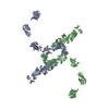 9ba4C M: map data used to model this data C: citing same article ( |
|---|---|
| Similar structure data |

-Links
 PDBj
PDBj


- Assembly
| Deposited unit | 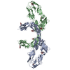
|
|---|---|
| 1 |
|
-Components
| #1: Protein | Mass: 65394.629 Da / Num. of mol.: 2 Source method: isolated from a genetically manipulated source Source: (gene. exp.) Homo sapiens (human) / Gene: CNTN2, AXT, TAG1, TAX1 / Production host:  Trichoplusia ni (cabbage looper) / References: UniProt: Q02246 Trichoplusia ni (cabbage looper) / References: UniProt: Q02246#2: Sugar | ChemComp-NAG /   Type: D-saccharide, beta linking / Mass: 221.208 Da / Num. of mol.: 10 / Source method: obtained synthetically / Formula: C8H15NO6 / Feature type: SUBJECT OF INVESTIGATION Type: D-saccharide, beta linking / Mass: 221.208 Da / Num. of mol.: 10 / Source method: obtained synthetically / Formula: C8H15NO6 / Feature type: SUBJECT OF INVESTIGATIONHas ligand of interest | Y | Has protein modification | Y | |
|---|
-Experimental details
-Experiment
| Experiment | Method: ELECTRON MICROSCOPY |
|---|---|
| EM experiment | Aggregation state: PARTICLE / 3D reconstruction method: single particle reconstruction |
- Sample preparation
| Component | Name: Contactin 2 Ig1-Ig6 / Type: ORGANELLE OR CELLULAR COMPONENT / Entity ID: #1 / Source: RECOMBINANT | |||||||||||||||
|---|---|---|---|---|---|---|---|---|---|---|---|---|---|---|---|---|
| Source (natural) | Organism: Homo sapiens (human) | |||||||||||||||
| Source (recombinant) | Organism: Trichoplusia ni (cabbage looper) | |||||||||||||||
| Buffer solution | pH: 8 / Details: 20 mM HEPES pH 8.0, 50 mM NaCl | |||||||||||||||
| Buffer component |
| |||||||||||||||
| Specimen | Conc.: 0.025 mg/ml / Embedding applied: NO / Shadowing applied: NO / Staining applied: NO / Vitrification applied: YES | |||||||||||||||
| Vitrification | Instrument: LEICA EM GP / Cryogen name: ETHANE / Humidity: 90 % / Chamber temperature: 281 K |
- Electron microscopy imaging
| Experimental equipment |  Model: Titan Krios / Image courtesy: FEI Company |
|---|---|
| Microscopy | Model: TFS KRIOS |
| Electron gun | Electron source:  FIELD EMISSION GUN / Accelerating voltage: 300 kV / Illumination mode: FLOOD BEAM FIELD EMISSION GUN / Accelerating voltage: 300 kV / Illumination mode: FLOOD BEAM |
| Electron lens | Mode: BRIGHT FIELD / Nominal magnification: 81000 X / Nominal defocus max: 1600 nm / Nominal defocus min: 600 nm |
| Specimen holder | Specimen holder model: FEI TITAN KRIOS AUTOGRID HOLDER |
| Image recording | Average exposure time: 7.39 sec. / Electron dose: 50 e/Å2 / Film or detector model: GATAN K3 (6k x 4k) / Num. of grids imaged: 1 / Num. of real images: 4297 |
| EM imaging optics | Energyfilter name: GIF Bioquantum / Energyfilter slit width: 20 eV |
- Processing
| EM software |
| ||||||||||||||||||||||||||||||||||||
|---|---|---|---|---|---|---|---|---|---|---|---|---|---|---|---|---|---|---|---|---|---|---|---|---|---|---|---|---|---|---|---|---|---|---|---|---|---|
| CTF correction | Type: PHASE FLIPPING AND AMPLITUDE CORRECTION | ||||||||||||||||||||||||||||||||||||
| 3D reconstruction | Resolution: 3.51 Å / Resolution method: FSC 0.143 CUT-OFF / Num. of particles: 98082 / Symmetry type: POINT | ||||||||||||||||||||||||||||||||||||
| Atomic model building | Protocol: FLEXIBLE FIT | ||||||||||||||||||||||||||||||||||||
| Refinement | Cross valid method: NONE Stereochemistry target values: GeoStd + Monomer Library + CDL v1.2 | ||||||||||||||||||||||||||||||||||||
| Displacement parameters | Biso mean: 100.14 Å2 | ||||||||||||||||||||||||||||||||||||
| Refine LS restraints |
|
