National Institutes of Health/National Institute of Mental Health (NIH/NIMH)
United States
Citation
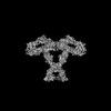
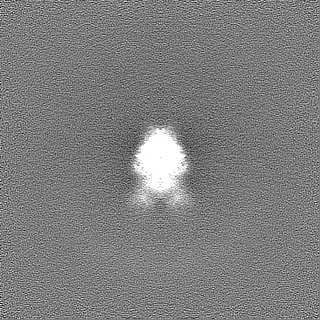
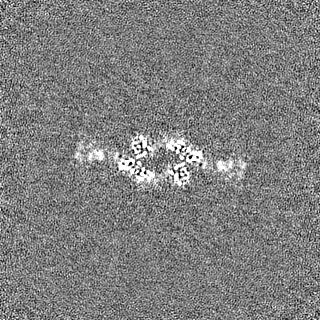
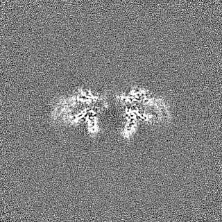
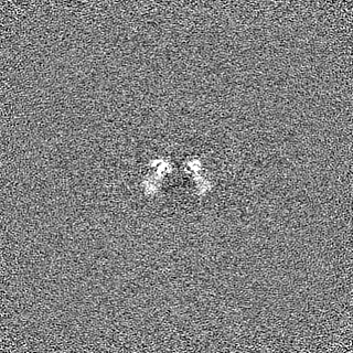
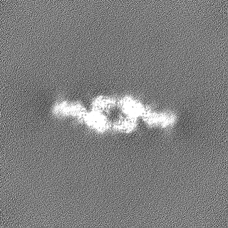
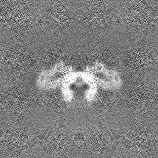
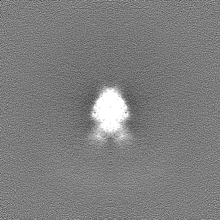
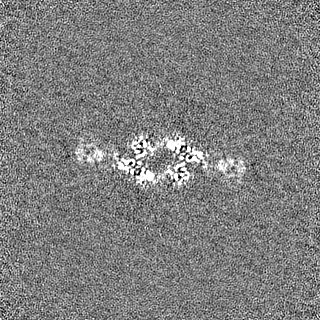
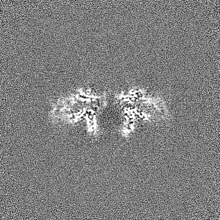
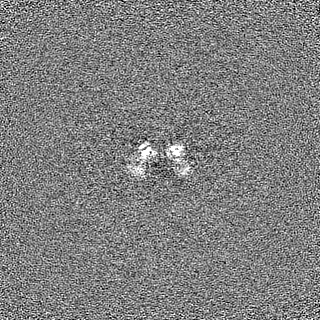
Journal: Structure / Year: 2024 Title: Molecular mechanism of contactin 2 homophilic interaction. Authors: Shanghua Fan / Jianfang Liu / Nicolas Chofflet / Aaron O Bailey / William K Russell / Ziqi Zhang / Hideto Takahashi / Gang Ren / Gabby Rudenko / Abstract: Contactin 2 (CNTN2) is a cell adhesion molecule involved in axon guidance, neuronal migration, and fasciculation. The ectodomains of CNTN1-CNTN6 are composed of six Ig domains (Ig1-Ig6) and four FN ...Contactin 2 (CNTN2) is a cell adhesion molecule involved in axon guidance, neuronal migration, and fasciculation. The ectodomains of CNTN1-CNTN6 are composed of six Ig domains (Ig1-Ig6) and four FN domains. Here, we show that CNTN2 forms transient homophilic interactions (K ∼200 nM). Cryo-EM structures of full-length CNTN2 and CNTN2_Ig1-Ig6 reveal a T-shaped homodimer formed by intertwined, parallel monomers. Unexpectedly, the horseshoe-shaped Ig1-Ig4 headpieces extend their Ig2-Ig3 tips outwards on either side of the homodimer, while Ig4, Ig5, Ig6, and the FN domains form a central stalk. Cross-linking mass spectrometry and cell-based binding assays confirm the 3D assembly of the CNTN2 homodimer. The interface mediating homodimer formation differs between CNTNs, as do the homophilic versus heterophilic interaction mechanisms. The CNTN family thus encodes a versatile molecular platform that supports a very diverse portfolio of protein interactions and that can be leveraged to strategically guide neural circuit development.
Cryogen name: ETHANE / Chamber humidity: 90 % / Chamber temperature: 281 K / Instrument: LEICA EM GP
-
Electron microscopy
Microscope
TFS KRIOS
Specialist optics
Energy filter - Name: GIF Bioquantum / Energy filter - Slit width: 20 eV
Image recording
Film or detector model: GATAN K3 (6k x 4k) / Number grids imaged: 1 / Number real images: 4297 / Average exposure time: 7.39 sec. / Average electron dose: 50.0 e/Å2
Electron beam
Acceleration voltage: 300 kV / Electron source: FIELD EMISSION GUN
In the structure databanks used in Yorodumi, some data are registered as the other names, "COVID-19 virus" and "2019-nCoV". Here are the details of the virus and the list of structure data.
Jan 31, 2019. EMDB accession codes are about to change! (news from PDBe EMDB page)
EMDB accession codes are about to change! (news from PDBe EMDB page)
The allocation of 4 digits for EMDB accession codes will soon come to an end. Whilst these codes will remain in use, new EMDB accession codes will include an additional digit and will expand incrementally as the available range of codes is exhausted. The current 4-digit format prefixed with “EMD-” (i.e. EMD-XXXX) will advance to a 5-digit format (i.e. EMD-XXXXX), and so on. It is currently estimated that the 4-digit codes will be depleted around Spring 2019, at which point the 5-digit format will come into force.
The EM Navigator/Yorodumi systems omit the EMD- prefix.
Related info.:Q: What is EMD? / ID/Accession-code notation in Yorodumi/EM Navigator
Yorodumi is a browser for structure data from EMDB, PDB, SASBDB, etc.
This page is also the successor to EM Navigator detail page, and also detail information page/front-end page for Omokage search.
The word "yorodu" (or yorozu) is an old Japanese word meaning "ten thousand". "mi" (miru) is to see.
Related info.:EMDB / PDB / SASBDB / Comparison of 3 databanks / Yorodumi Search / Aug 31, 2016. New EM Navigator & Yorodumi / Yorodumi Papers / Jmol/JSmol / Function and homology information / Changes in new EM Navigator and Yorodumi
 Movie
Movie Controller


 Open data
 Basic information
 Map data
 Sample
 Keywords
 Function and homology information
 Homo sapiens (human)
 Authors
 United States, 1 items
  Citation
 Structure visualization
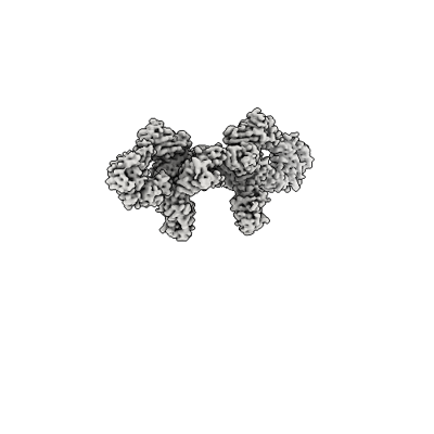
 Downloads & links
 emd_44396.png
 http://ftp.pdbj.org/pub/emdb/structures/EMD-44396
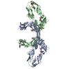


 Z (Sec.)
 Y (Row.)
 X (Col.)


 Sample components
 Trichoplusia ni (cabbage looper)
 Processing
 Electron microscopy
 FIELD EMISSION GUN