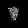
 ムービー
ムービー コントローラー


+ データを開く

- 基本情報
基本情報
| 登録情報 | データベース: PDB / ID: 9b73 | ||||||
|---|---|---|---|---|---|---|---|
| タイトル | Cryo-EM structure of the desensitised ATP-bound human P2X1 receptor | ||||||
 要素 要素 | P2X purinoceptor 1 | ||||||
 キーワード キーワード | MEMBRANE PROTEIN / Ion channel / trimer / ATP-bound / desensitised | ||||||
| 機能・相同性 |  機能・相同性情報 機能・相同性情報Platelet homeostasis / insemination / positive regulation of calcium ion import across plasma membrane / extracellularly ATP-gated monoatomic cation channel activity / purinergic nucleotide receptor activity / suramin binding / regulation of vascular associated smooth muscle contraction / ligand-gated calcium channel activity / serotonin secretion by platelet / Elevation of cytosolic Ca2+ levels ...Platelet homeostasis / insemination / positive regulation of calcium ion import across plasma membrane / extracellularly ATP-gated monoatomic cation channel activity / purinergic nucleotide receptor activity / suramin binding / regulation of vascular associated smooth muscle contraction / ligand-gated calcium channel activity / serotonin secretion by platelet / Elevation of cytosolic Ca2+ levels / regulation of presynaptic cytosolic calcium ion concentration / ceramide biosynthetic process / response to ATP / regulation of synaptic vesicle exocytosis / neuronal action potential / monoatomic cation channel activity / specific granule membrane / monoatomic ion transport / presynaptic active zone membrane / secretory granule membrane / synaptic transmission, glutamatergic / calcium ion transmembrane transport / platelet activation / regulation of blood pressure / postsynaptic membrane / membrane raft / external side of plasma membrane / apoptotic process / Neutrophil degranulation / protein-containing complex binding / glutamatergic synapse / signal transduction / protein-containing complex / ATP binding / identical protein binding / plasma membrane 類似検索 - 分子機能 | ||||||
| 生物種 |  Homo sapiens (ヒト) Homo sapiens (ヒト) | ||||||
| 手法 | 電子顕微鏡法 / 単粒子再構成法 / クライオ電子顕微鏡法 / 解像度: 1.96 Å | ||||||
 データ登録者 データ登録者 | Felix, M.B. / Alisa, G. / Hariprasad, V. / Jesse, I.M. / David, M.T. | ||||||
| 資金援助 |  オーストラリア, 1件 オーストラリア, 1件
| ||||||
 引用 引用 | ジャーナル: Nat Commun / 年: 2024 タイトル: Structural insights into the human P2X1 receptor and ligand interactions. 著者: Felix M Bennetts / Hariprasad Venugopal / Alisa Glukhova / Jesse I Mobbs / Sabatino Ventura / David M Thal / 要旨: The P2X1 receptor is a trimeric ligand-gated ion channel that plays an important role in urogenital and immune functions, offering the potential for new drug treatments. However, progress in this ...The P2X1 receptor is a trimeric ligand-gated ion channel that plays an important role in urogenital and immune functions, offering the potential for new drug treatments. However, progress in this area has been hindered by limited structural information and a lack of well-characterised tool compounds. In this study, we employ cryogenic electron microscopy (cryo-EM) to elucidate the structures of the P2X1 receptor in an ATP-bound desensitised state and an NF449-bound closed state. NF449, a potent P2X1 receptor antagonist, engages the receptor distinctively, while ATP, the endogenous ligand, binds in a manner consistent with other P2X receptors. To explore the molecular basis of receptor inhibition, activation, and ligand interactions, key residues involved in ligand and metal ion binding were mutated. Radioligand binding assays with [H]-α,β-methylene ATP and intracellular calcium ion influx assays were used to evaluate the effects of these mutations. These experiments validate key ligand-receptor interactions and identify conserved and non-conserved residues critical for ligand binding or receptor modulation. This research expands our understanding of the P2X1 receptor structure at a molecular level and opens new avenues for in silico drug design targeting the P2X1 receptor. | ||||||
| 履歴 |
|
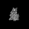
- 構造の表示
構造の表示
| 構造ビューア | 分子: MolmilJmol/JSmol |
|---|
- ダウンロードとリンク
-ダウンロード
| PDBx/mmCIF形式 | 9b73.cif.gz | 245.6 KB | 表示 | PDBx/mmCIF形式 |
|---|---|---|---|---|
| PDB形式 | pdb9b73.ent.gz | 表示 | PDB形式 | |
| PDBx/mmJSON形式 | 9b73.json.gz | ツリー表示 | PDBx/mmJSON形式 | |
| その他 |  その他のダウンロード その他のダウンロード |
-検証レポート
| 文書・要旨 | 9b73_validation.pdf.gz | 1.3 MB | 表示 | wwPDB検証レポート |
|---|---|---|---|---|
| 文書・詳細版 | 9b73_full_validation.pdf.gz | 1.4 MB | 表示 | |
| XML形式データ | 9b73_validation.xml.gz | 41.9 KB | 表示 | |
| CIF形式データ | 9b73_validation.cif.gz | 58.8 KB | 表示 | |
| アーカイブディレクトリ | https://data.pdbj.org/pub/pdb/validation_reports/b7/9b73ftp://data.pdbj.org/pub/pdb/validation_reports/b7/9b73 | HTTPS FTP |
-関連構造データ

-リンク
 PDBj
PDBj- 集合体
集合体
| 登録構造単位 | 
|
|---|---|
| 1 |
|
-要素
| #1: タンパク質 | 分子量: 45038.957 Da / 分子数: 3 / 由来タイプ: 組換発現 / 由来: (組換発現) Homo sapiens (ヒト) / 遺伝子: P2RX1, P2X1発現宿主:   Spodoptera frugiperda (ツマジロクサヨトウ) Spodoptera frugiperda (ツマジロクサヨトウ)参照: UniProt: P51575 #2: 化合物 |   分子量: 507.181 Da / 分子数: 3 / 由来タイプ: 合成 / 式: C10H16N5O13P3 / タイプ: SUBJECT OF INVESTIGATION / コメント: ATP, エネルギー貯蔵分子*YM 分子量: 507.181 Da / 分子数: 3 / 由来タイプ: 合成 / 式: C10H16N5O13P3 / タイプ: SUBJECT OF INVESTIGATION / コメント: ATP, エネルギー貯蔵分子*YM#3: 糖 | ChemComp-NAG /   タイプ: D-saccharide, beta linking / 分子量: 221.208 Da / 分子数: 9 / 由来タイプ: 合成 / 式: C8H15NO6 タイプ: D-saccharide, beta linking / 分子量: 221.208 Da / 分子数: 9 / 由来タイプ: 合成 / 式: C8H15NO6#4: 化合物 |   分子量: 24.305 Da / 分子数: 3 / 由来タイプ: 合成 / 式: Mg 分子量: 24.305 Da / 分子数: 3 / 由来タイプ: 合成 / 式: Mg#5: 水 | ChemComp-HOH / |  分子量: 18.015 Da / 分子数: 66 / 由来タイプ: 天然 / 式: H2O 分子量: 18.015 Da / 分子数: 66 / 由来タイプ: 天然 / 式: H2O研究の焦点であるリガンドがあるか | Y | Has protein modification | Y | |
|---|
-実験情報
-実験
| 実験 | 手法: 電子顕微鏡法 |
|---|---|
| EM実験 | 試料の集合状態: PARTICLE / 3次元再構成法: 単粒子再構成法 |
- 試料調製
試料調製
| 構成要素 | 名称: P2X1 receptor trimer / タイプ: COMPLEX / Entity ID: #1 / 由来: RECOMBINANT | ||||||||||||||||||||||||||||||||||||||||
|---|---|---|---|---|---|---|---|---|---|---|---|---|---|---|---|---|---|---|---|---|---|---|---|---|---|---|---|---|---|---|---|---|---|---|---|---|---|---|---|---|---|
| 分子量 | 値: 0.134940 MDa / 実験値: NO | ||||||||||||||||||||||||||||||||||||||||
| 由来(天然) | 生物種: Homo sapiens (ヒト) | ||||||||||||||||||||||||||||||||||||||||
| 由来(組換発現) | 生物種: Spodoptera frugiperda (ツマジロクサヨトウ) | ||||||||||||||||||||||||||||||||||||||||
| 緩衝液 | pH: 8 詳細: The buffer consists of 50 mM Tris (pH 8), 100 mM NaCl, 0.01% LMNG, and 0.0006% CHS supplemented with 1 mM ATP and 1 mM MgCl2 overnight before vitrification. Additionally, 0.4 mM or 1.2 mM of ...詳細: The buffer consists of 50 mM Tris (pH 8), 100 mM NaCl, 0.01% LMNG, and 0.0006% CHS supplemented with 1 mM ATP and 1 mM MgCl2 overnight before vitrification. Additionally, 0.4 mM or 1.2 mM of fluorinated Fos-Choline-8 (fluor-FC8) was added just one minute prior to vitrification. | ||||||||||||||||||||||||||||||||||||||||
| 緩衝液成分 |
| ||||||||||||||||||||||||||||||||||||||||
| 試料 | 濃度: 19 mg/ml / 包埋: NO / シャドウイング: NO / 染色: NO / 凍結: YES | ||||||||||||||||||||||||||||||||||||||||
| 試料支持 | 詳細: Model used: Glow Discharge Peelco Easyglow / グリッドの材料: GOLD / グリッドのサイズ: 300 divisions/in. / グリッドのタイプ: UltrAuFoil R1.2/1.3 | ||||||||||||||||||||||||||||||||||||||||
| 急速凍結 | 装置: FEI VITROBOT MARK III / 凍結剤: ETHANE / 湿度: 100 % / 凍結前の試料温度: 277 K 詳細: Grids were frozen in liquid ethane using a Vitrobot Mark III operated at 4 degrees Celsius and 100% humidity with 12 blot force and 2 seconds blot time. |
- 電子顕微鏡撮影
| 実験機器 |  モデル: Titan Krios / 画像提供: FEI Company |
|---|---|
| 顕微鏡 | モデル: TFS KRIOS 詳細: Initial grid screening was conducted using a 200kV TFS Artica cryo-electron microscope prior to imaging on a Titan Krios cryo-electron microscope. |
| 電子銃 | 電子線源:  FIELD EMISSION GUN / 加速電圧: 300 kV / 照射モード: FLOOD BEAM FIELD EMISSION GUN / 加速電圧: 300 kV / 照射モード: FLOOD BEAM |
| 電子レンズ | モード: BRIGHT FIELD / 倍率(公称値): 105000 X / 倍率(補正後): 105000 X / 最大 デフォーカス(公称値): 1500 nm / 最小 デフォーカス(公称値): 500 nm / Cs: 2.7 mm |
| 試料ホルダ | 凍結剤: NITROGEN 試料ホルダーモデル: FEI TITAN KRIOS AUTOGRID HOLDER |
| 撮影 | 平均露光時間: 1 sec. / 電子線照射量: 60 e/Å2 / フィルム・検出器のモデル: GATAN K3 (6k x 4k) / 撮影したグリッド数: 2 / 実像数: 10756 詳細: 4578 images were collected on the grid containing P2X1 receptor supplemented with a higher concentration of fluor-FC8 (1.2 mM), while 6178 images were collected on the grid with a lower ...詳細: 4578 images were collected on the grid containing P2X1 receptor supplemented with a higher concentration of fluor-FC8 (1.2 mM), while 6178 images were collected on the grid with a lower concentration of fluor-FC8 (0.4 mM) |
- 解析
解析
| EMソフトウェア |
| |||||||||||||||||||||||||||||||||||||||||||||
|---|---|---|---|---|---|---|---|---|---|---|---|---|---|---|---|---|---|---|---|---|---|---|---|---|---|---|---|---|---|---|---|---|---|---|---|---|---|---|---|---|---|---|---|---|---|---|
| CTF補正 | 詳細: CTF correction was performed on the initial set of motion-corrected images, followed by subsequent local CTF corrections on the final subset of particles. タイプ: PHASE FLIPPING AND AMPLITUDE CORRECTION | |||||||||||||||||||||||||||||||||||||||||||||
| 粒子像の選択 | 選択した粒子像数: 5312810 詳細: Initially, 2,565,511 particles were picked for the higher concentration fluor-FC8 P2X1 receptor sample, and 2,747,299 particles were picked for the lower concentration sample. | |||||||||||||||||||||||||||||||||||||||||||||
| 対称性 | 点対称性: C3 (3回回転対称) | |||||||||||||||||||||||||||||||||||||||||||||
| 3次元再構成 | 解像度: 1.96 Å / 解像度の算出法: FSC 0.143 CUT-OFF / 粒子像の数: 481309 / アルゴリズム: FOURIER SPACE / クラス平均像の数: 1 / 対称性のタイプ: POINT | |||||||||||||||||||||||||||||||||||||||||||||
| 原子モデル構築 | B value: 60.7 / プロトコル: RIGID BODY FIT / 空間: REAL 詳細: Initial fitting was performed in Chimerax and then refinements were performed in Coot and Phenix. | |||||||||||||||||||||||||||||||||||||||||||||
| 原子モデル構築 | Accession code: P51575 / Chain residue range: 1-399 / 詳細: Full human P2X1 receptor trimer / Source name: AlphaFold / タイプ: in silico model | |||||||||||||||||||||||||||||||||||||||||||||
| 精密化 | 交差検証法: NONE 立体化学のターゲット値: GeoStd + Monomer Library + CDL v1.2 | |||||||||||||||||||||||||||||||||||||||||||||
| 原子変位パラメータ | Biso mean: 92.01 Å2 | |||||||||||||||||||||||||||||||||||||||||||||
| 拘束条件 |
|
