 Movie
Movie Controller

[English] 日本語
 Yorodumi
- EMDB-44299: Cryo-EM structure of the desensitised ATP-bound human P2X1 receptor -
+ Open data

- Basic information
| Entry | 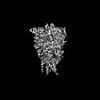 | |||||||||
|---|---|---|---|---|---|---|---|---|---|---|
| Title | Cryo-EM structure of the desensitised ATP-bound human P2X1 receptor | |||||||||
 Map data Map data | Map of the P2X1 receptor generated in Cryosparc. (J609 homogenous refinement job) | |||||||||
 Sample Sample |
| |||||||||
 Keywords Keywords | Ion channel / trimer / ATP-bound / desensitised / MEMBRANE PROTEIN | |||||||||
| Function / homology |  Function and homology information Function and homology informationinsemination / Platelet homeostasis / positive regulation of calcium ion import across plasma membrane / suramin binding / serotonin secretion by platelet / regulation of vascular associated smooth muscle contraction / extracellularly ATP-gated monoatomic cation channel activity / purinergic nucleotide receptor activity / regulation of presynaptic cytosolic calcium ion concentration / Elevation of cytosolic Ca2+ levels ...insemination / Platelet homeostasis / positive regulation of calcium ion import across plasma membrane / suramin binding / serotonin secretion by platelet / regulation of vascular associated smooth muscle contraction / extracellularly ATP-gated monoatomic cation channel activity / purinergic nucleotide receptor activity / regulation of presynaptic cytosolic calcium ion concentration / Elevation of cytosolic Ca2+ levels / ceramide biosynthetic process / ligand-gated calcium channel activity / regulation of synaptic vesicle exocytosis / response to ATP / specific granule membrane / monoatomic cation channel activity / monoatomic ion transport / neuronal action potential / presynaptic active zone membrane / secretory granule membrane / synaptic transmission, glutamatergic / platelet activation / regulation of blood pressure / calcium ion transmembrane transport / postsynaptic membrane / membrane raft / external side of plasma membrane / apoptotic process / Neutrophil degranulation / protein-containing complex binding / glutamatergic synapse / signal transduction / protein-containing complex / ATP binding / identical protein binding / plasma membrane Similarity search - Function | |||||||||
| Biological species |  Homo sapiens (human) Homo sapiens (human) | |||||||||
| Method | single particle reconstruction / cryo EM / Resolution: 1.96 Å | |||||||||
 Authors Authors | Felix MB / Alisa G / Hariprasad V / Jesse IM / David MT | |||||||||
| Funding support |  Australia, 1 items Australia, 1 items
| |||||||||
 Citation Citation | Journal: Nat Commun / Year: 2024 Title: Structural insights into the human P2X1 receptor and ligand interactions. Authors: Felix M Bennetts / Hariprasad Venugopal / Alisa Glukhova / Jesse I Mobbs / Sabatino Ventura / David M Thal / Abstract: The P2X1 receptor is a trimeric ligand-gated ion channel that plays an important role in urogenital and immune functions, offering the potential for new drug treatments. However, progress in this ...The P2X1 receptor is a trimeric ligand-gated ion channel that plays an important role in urogenital and immune functions, offering the potential for new drug treatments. However, progress in this area has been hindered by limited structural information and a lack of well-characterised tool compounds. In this study, we employ cryogenic electron microscopy (cryo-EM) to elucidate the structures of the P2X1 receptor in an ATP-bound desensitised state and an NF449-bound closed state. NF449, a potent P2X1 receptor antagonist, engages the receptor distinctively, while ATP, the endogenous ligand, binds in a manner consistent with other P2X receptors. To explore the molecular basis of receptor inhibition, activation, and ligand interactions, key residues involved in ligand and metal ion binding were mutated. Radioligand binding assays with [H]-α,β-methylene ATP and intracellular calcium ion influx assays were used to evaluate the effects of these mutations. These experiments validate key ligand-receptor interactions and identify conserved and non-conserved residues critical for ligand binding or receptor modulation. This research expands our understanding of the P2X1 receptor structure at a molecular level and opens new avenues for in silico drug design targeting the P2X1 receptor. | |||||||||
| History |
|
- Structure visualization
| Supplemental images |
|---|

- Downloads & links
-EMDB archive
| Map data | emd_44299.map.gz | 26.4 MB | EMDB map data format | |
|---|---|---|---|---|
| Header (meta data) | emd-44299-v30.xmlemd-44299.xml | 24.9 KB 24.9 KB | Display Display | EMDB header |
| FSC (resolution estimation) | emd_44299_fsc.xml | 7.8 KB | Display | FSC data file |
| Images |  emd_44299.png emd_44299.png | 65.4 KB | ||
| Masks | emd_44299_msk_1.map | 52.7 MB | Mask map | |
| Filedesc metadata | emd-44299.cif.gz | 7.5 KB | ||
| Others | emd_44299_additional_1.map.gzemd_44299_half_map_1.map.gzemd_44299_half_map_2.map.gz | 49.3 MB 48.9 MB 48.9 MB | ||
| Archive directory |  http://ftp.pdbj.org/pub/emdb/structures/EMD-44299ftp://ftp.pdbj.org/pub/emdb/structures/EMD-44299 http://ftp.pdbj.org/pub/emdb/structures/EMD-44299ftp://ftp.pdbj.org/pub/emdb/structures/EMD-44299 | HTTPS FTP |
-Related structure data
| Related structure data | 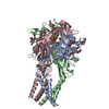 9b73MC  9b95C M: atomic model generated by this map C: citing same article ( |
|---|---|
| Similar structure data |
-Links
| EMDB pages | EMDB (EBI/PDBe) / EMDataResource |
|---|
-Map
| File | Download / File: emd_44299.map.gz / Format: CCP4 / Size: 52.7 MB / Type: IMAGE STORED AS FLOATING POINT NUMBER (4 BYTES) | ||||||||||||||||||||||||||||||||||||
|---|---|---|---|---|---|---|---|---|---|---|---|---|---|---|---|---|---|---|---|---|---|---|---|---|---|---|---|---|---|---|---|---|---|---|---|---|---|
| Annotation | Map of the P2X1 receptor generated in Cryosparc. (J609 homogenous refinement job) | ||||||||||||||||||||||||||||||||||||
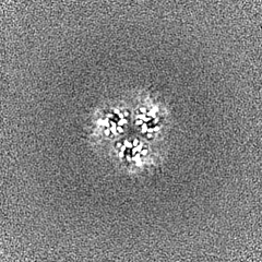
| Projections & slices | Image control
Images are generated by Spider. | ||||||||||||||||||||||||||||||||||||
| Voxel size | X=Y=Z: 0.82 Å | ||||||||||||||||||||||||||||||||||||
| Density |
| ||||||||||||||||||||||||||||||||||||
| Symmetry | Space group: 1 | ||||||||||||||||||||||||||||||||||||
| Details | EMDB XML:
|
 Z (Sec.)
 Y (Row.)
 X (Col.)


-Supplemental data
-Mask #1
| File | emd_44299_msk_1.map | ||||||||||||
|---|---|---|---|---|---|---|---|---|---|---|---|---|---|
| Projections & Slices |
| ||||||||||||
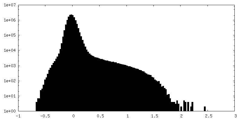
| Density Histograms |


-Additional map: Sharp map of the P2X1 receptor generated in...
| File | emd_44299_additional_1.map | ||||||||||||
|---|---|---|---|---|---|---|---|---|---|---|---|---|---|
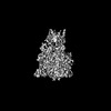
| Annotation | Sharp map of the P2X1 receptor generated in Phenix. (Generated from original cryo-EM map) | ||||||||||||
| Projections & Slices |
| ||||||||||||
| Density Histograms |


-Half map: Half map of the P2X1 receptor generated in...
| File | emd_44299_half_map_1.map | ||||||||||||
|---|---|---|---|---|---|---|---|---|---|---|---|---|---|
| Annotation | Half map of the P2X1 receptor generated in Cryosparc. (J609 homogenous refinement job) | ||||||||||||
| Projections & Slices |
| ||||||||||||
| Density Histograms |


-Half map: Half map of the P2X1 receptor generated in...
| File | emd_44299_half_map_2.map | ||||||||||||
|---|---|---|---|---|---|---|---|---|---|---|---|---|---|
| Annotation | Half map of the P2X1 receptor generated in Cryosparc. (J609 homogenous refinement job) | ||||||||||||
| Projections & Slices |
| ||||||||||||
| Density Histograms |


- Sample components
-Entire : P2X1 receptor trimer
| Entire | Name: P2X1 receptor trimer |
|---|---|
| Components |
|
-Supramolecule #1: P2X1 receptor trimer
| Supramolecule | Name: P2X1 receptor trimer / type: complex / ID: 1 / Parent: 0 / Macromolecule list: #1 |
|---|---|
| Source (natural) | Organism: Homo sapiens (human) |
| Molecular weight | Theoretical: 134.94 KDa |
-Macromolecule #1: P2X purinoceptor 1
| Macromolecule | Name: P2X purinoceptor 1 / type: protein_or_peptide / ID: 1 / Number of copies: 3 / Enantiomer: LEVO |
|---|---|
| Source (natural) | Organism: Homo sapiens (human) |
| Molecular weight | Theoretical: 45.038957 KDa |
| Recombinant expression | Organism:   Spodoptera frugiperda (fall armyworm) Spodoptera frugiperda (fall armyworm) |
| Sequence | String: MARRFQEELA AFLFEYDTPR MVLVRNKKVG VIFRLIQLVV LVYVIGWVFL YEKGYQTSSG LISSVSVKLK GLAVTQLPGL GPQVWDVAD YVFPAQGDNS FVVMTNFIVT PKQTQGYCAE HPEGGICKED SGCTPGKAKR KAQGIRTGKC VAFNDTVKTC E IFGWCPVE ...String: MARRFQEELA AFLFEYDTPR MVLVRNKKVG VIFRLIQLVV LVYVIGWVFL YEKGYQTSSG LISSVSVKLK GLAVTQLPGL GPQVWDVAD YVFPAQGDNS FVVMTNFIVT PKQTQGYCAE HPEGGICKED SGCTPGKAKR KAQGIRTGKC VAFNDTVKTC E IFGWCPVE VDDDIPRPAL LREAENFTLF IKNSISFPRF KVNRRNLVEE VNAAHMKTCL FHKTLHPLCP VFQLGYVVQE SG QNFSTLA EKGGVVGITI DWHCDLDWHV RHCRPIYEFH GLYEEKNLSP GFNFRFARHF VENGTNYRHL FKVFGIRFDI LVD GKAGKF DIIPTMTTIG SGIGIFGVAT VLCDLLLLHI LPKRHYYKQK KFKYAEDMGP GAAERDLAAT SSTLGLQENM RTS UniProtKB: P2X purinoceptor 1 |
-Macromolecule #2: ADENOSINE-5'-TRIPHOSPHATE
| Macromolecule | Name: ADENOSINE-5'-TRIPHOSPHATE / type: ligand / ID: 2 / Number of copies: 3 / Formula: ATP |
|---|---|
| Molecular weight | Theoretical: 507.181 Da |
| Chemical component information |  ChemComp-ATP: |
-Macromolecule #3: 2-acetamido-2-deoxy-beta-D-glucopyranose
| Macromolecule | Name: 2-acetamido-2-deoxy-beta-D-glucopyranose / type: ligand / ID: 3 / Number of copies: 9 / Formula: NAG |
|---|---|
| Molecular weight | Theoretical: 221.208 Da |
| Chemical component information |  ChemComp-NAG: |
-Macromolecule #4: MAGNESIUM ION
| Macromolecule | Name: MAGNESIUM ION / type: ligand / ID: 4 / Number of copies: 3 / Formula: MG |
|---|---|
| Molecular weight | Theoretical: 24.305 Da |
-Macromolecule #5: water
| Macromolecule | Name: water / type: ligand / ID: 5 / Number of copies: 66 / Formula: HOH |
|---|---|
| Molecular weight | Theoretical: 18.015 Da |
| Chemical component information |  ChemComp-HOH: |
-Experimental details
-Structure determination
| Method | cryo EM |
|---|---|
 Processing Processing | single particle reconstruction |
| Aggregation state | particle |
-Sample preparation
| Concentration | 19 mg/mL | ||||||||||||||||||||||||
|---|---|---|---|---|---|---|---|---|---|---|---|---|---|---|---|---|---|---|---|---|---|---|---|---|---|
| Buffer | pH: 8 Component:
Details: The buffer consists of 50 mM Tris (pH 8), 100 mM NaCl, 0.01% LMNG, and 0.0006% CHS supplemented with 1 mM ATP and 1 mM MgCl2 overnight before vitrification. Additionally, 0.4 mM or 1.2 mM of ...Details: The buffer consists of 50 mM Tris (pH 8), 100 mM NaCl, 0.01% LMNG, and 0.0006% CHS supplemented with 1 mM ATP and 1 mM MgCl2 overnight before vitrification. Additionally, 0.4 mM or 1.2 mM of fluorinated Fos-Choline-8 (fluor-FC8) was added just one minute prior to vitrification. | ||||||||||||||||||||||||
| Grid | Model: UltrAuFoil R1.2/1.3 / Material: GOLD / Mesh: 300 / Support film - Material: GOLD / Support film - topology: HOLEY ARRAY / Support film - Film thickness: 50 / Pretreatment - Type: GLOW DISCHARGE / Pretreatment - Time: 180 sec. / Pretreatment - Atmosphere: OTHER / Details: Model used: Glow Discharge Peelco Easyglow | ||||||||||||||||||||||||
| Vitrification | Cryogen name: ETHANE / Chamber humidity: 100 % / Chamber temperature: 277 K / Instrument: FEI VITROBOT MARK III Details: Grids were frozen in liquid ethane using a Vitrobot Mark III operated at 4 degrees Celsius and 100% humidity with 12 blot force and 2 seconds blot time.. |
- Electron microscopy
| Microscope | TFS KRIOS |
|---|---|
| Details | Initial grid screening was conducted using a 200kV TFS Artica cryo-electron microscope prior to imaging on a Titan Krios cryo-electron microscope. |
| Image recording | Film or detector model: GATAN K3 (6k x 4k) / Number grids imaged: 2 / Number real images: 10756 / Average exposure time: 1.0 sec. / Average electron dose: 60.0 e/Å2 Details: 4578 images were collected on the grid containing P2X1 receptor supplemented with a higher concentration of fluor-FC8 (1.2 mM), while 6178 images were collected on the grid with a lower ...Details: 4578 images were collected on the grid containing P2X1 receptor supplemented with a higher concentration of fluor-FC8 (1.2 mM), while 6178 images were collected on the grid with a lower concentration of fluor-FC8 (0.4 mM) |
| Electron beam | Acceleration voltage: 300 kV / Electron source:  FIELD EMISSION GUN FIELD EMISSION GUN |
| Electron optics | Calibrated magnification: 105000 / Illumination mode: FLOOD BEAM / Imaging mode: BRIGHT FIELD / Cs: 2.7 mm / Nominal defocus max: 1.5 µm / Nominal defocus min: 0.5 µm / Nominal magnification: 105000 |
| Sample stage | Specimen holder model: FEI TITAN KRIOS AUTOGRID HOLDER / Cooling holder cryogen: NITROGEN |
| Experimental equipment |  Model: Titan Krios / Image courtesy: FEI Company |
+Image processing

-Atomic model buiding 1
| Initial model | PDB ID: Chain - Residue range: 1-399 / Chain - Source name: AlphaFold / Chain - Initial model type: in silico model / Details: Full human P2X1 receptor trimer |
|---|---|
| Details | Initial fitting was performed in Chimerax and then refinements were performed in Coot and Phenix. |
| Refinement | Space: REAL / Protocol: RIGID BODY FIT / Overall B value: 60.7 |
| Output model | PDB-9b73: |

