 Movie
Movie Controller

[English] 日本語
 Yorodumi
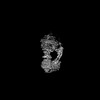
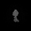
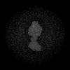
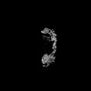
- PDB-9b0x: Artemia franciscana ATP synthase state 2 (composite structure), pH 7.0 -
+ Open data

- Basic information
| Entry | Database: PDB / ID: 9b0x | |||||||||||||||||||||||||||
|---|---|---|---|---|---|---|---|---|---|---|---|---|---|---|---|---|---|---|---|---|---|---|---|---|---|---|---|---|
| Title | Artemia franciscana ATP synthase state 2 (composite structure), pH 7.0 | |||||||||||||||||||||||||||
 Components Components | (ATP synthase ...) x 17 | |||||||||||||||||||||||||||
 Keywords Keywords | MEMBRANE PROTEIN / ATP synthesis / Complex V / mitochondria / oxidative-phosphorylation | |||||||||||||||||||||||||||
| Function / homology |  Function and homology information Function and homology informationATP biosynthetic process / proton-transporting ATP synthase complex / proton-transporting ATP synthase activity, rotational mechanism / proton transmembrane transport / mitochondrial membrane / mitochondrial inner membrane Similarity search - Function | |||||||||||||||||||||||||||
| Biological species |  Artemia franciscana (arthropod) Artemia franciscana (arthropod) | |||||||||||||||||||||||||||
| Method | ELECTRON MICROSCOPY / single particle reconstruction / cryo EM / Resolution: 2.6 Å | |||||||||||||||||||||||||||
 Authors Authors | Mnatsakanyan, N. / Mello, J.F.R. | |||||||||||||||||||||||||||
| Funding support |  United States, 2items United States, 2items
| |||||||||||||||||||||||||||
 Citation Citation | Journal: Cell Death Differ / Year: 2025 Title: Cryo-EM structure of the brine shrimp mitochondrial ATP synthase suggests an inactivation mechanism for the ATP synthase leak channel. Authors: Amrendra Kumar / Juliana da Fonseca Rezende E Mello / Yangyu Wu / Daniel Morris / Ikram Mezghani / Erin Smith / Stephane Rombauts / Peter Bossier / Juno Krahn / Fred J Sigworth / Nelli Mnatsakanyan /  Abstract: Mammalian mitochondria undergo Ca-induced and cyclosporinA (CsA)-regulated permeability transition (mPT) by activating the mitochondrial permeability transition pore (mPTP) situated in mitochondrial ...Mammalian mitochondria undergo Ca-induced and cyclosporinA (CsA)-regulated permeability transition (mPT) by activating the mitochondrial permeability transition pore (mPTP) situated in mitochondrial inner membranes. Ca-induced prolonged openings of mPTP under certain pathological conditions result in mitochondrial swelling and rupture of the outer membrane, leading to mitochondrial dysfunction and cell death. While the exact molecular composition and structure of mPTP remain unknown, mammalian ATP synthase was reported to form voltage and Ca-activated leak channels involved in mPT. Unlike in mammals, mitochondria of the crustacean Artemia franciscana have the ability to accumulate large amounts of Ca without undergoing the mPT. Here, we performed structural and functional analysis of A. franciscana ATP synthase to study the molecular mechanism of mPTP inhibition in this organism. We found that the channel formed by the A. franciscana ATP synthase dwells predominantly in its inactive state and is insensitive to Ca, in contrast to porcine heart ATP synthase. Single-particle cryo-electron microscopy (cryo-EM) analysis revealed distinct structural features in A. franciscana ATP synthase compared with mammals. The stronger density of the e-subunit C-terminal region and its enhanced interaction with the c-ring were found in A. franciscana ATP synthase. These data suggest an inactivation mechanism of the ATP synthase leak channel and its possible contribution to the lack of mPT in this organism. | |||||||||||||||||||||||||||
| History |
|
- Structure visualization
| Structure viewer | Molecule: MolmilJmol/JSmol |
|---|
- Downloads & links
-Download
| PDBx/mmCIF format | 9b0x.cif.gz | 997.9 KB | Display | PDBx/mmCIF format |
|---|---|---|---|---|
| PDB format | pdb9b0x.ent.gz | Display | PDB format | |
| PDBx/mmJSON format | 9b0x.json.gz | Tree view | PDBx/mmJSON format | |
| Others |  Other downloads Other downloads |
-Validation report
| Arichive directory | https://data.pdbj.org/pub/pdb/validation_reports/b0/9b0xftp://data.pdbj.org/pub/pdb/validation_reports/b0/9b0x | HTTPS FTP |
|---|
-Related structure data
| Related structure data | 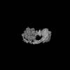 44061MC 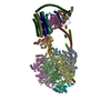 9b3jC 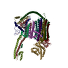 9bpgC M: map data used to model this data C: citing same article ( |
|---|---|
| Similar structure data |

-Links
 PDBj
PDBj


- Assembly
| Deposited unit | 
|
|---|---|
| 1 |
|
-Components
-ATP synthase ... , 17 types, 28 molecules 12345678ABCDEFGHIJKLMNOPQRST
| #1: Protein | Mass: 13250.408 Da / Num. of mol.: 8 / Source method: isolated from a natural source / Source: (natural) Artemia franciscana (arthropod)#2: Protein | Mass: 59706.500 Da / Num. of mol.: 3 / Source method: isolated from a natural source / Source: (natural) Artemia franciscana (arthropod)#3: Protein | Mass: 55884.770 Da / Num. of mol.: 3 / Source method: isolated from a natural source / Source: (natural) Artemia franciscana (arthropod)#4: Protein | | Mass: 31976.818 Da / Num. of mol.: 1 / Source method: isolated from a natural source / Source: (natural) Artemia franciscana (arthropod)#5: Protein | | Mass: 17565.455 Da / Num. of mol.: 1 / Source method: isolated from a natural source / Source: (natural) Artemia franciscana (arthropod)#6: Protein | | Mass: 7464.596 Da / Num. of mol.: 1 / Source method: isolated from a natural source / Source: (natural) Artemia franciscana (arthropod)#7: Protein | | Mass: 11974.477 Da / Num. of mol.: 1 / Source method: isolated from a natural source / Source: (natural) Artemia franciscana (arthropod)#8: Protein | | Mass: 29944.416 Da / Num. of mol.: 1 / Source method: isolated from a natural source / Source: (natural) Artemia franciscana (arthropod)#9: Protein | | Mass: 11140.879 Da / Num. of mol.: 1 / Source method: isolated from a natural source / Source: (natural) Artemia franciscana (arthropod)#10: Protein | | Mass: 25422.525 Da / Num. of mol.: 1 / Source method: isolated from a natural source / Source: (natural) Artemia franciscana (arthropod)#11: Protein | | Mass: 24422.471 Da / Num. of mol.: 1 / Source method: isolated from a natural source / Source: (natural) Artemia franciscana (arthropod) / References: UniProt: Q37708#12: Protein | | Mass: 22563.432 Da / Num. of mol.: 1 / Source method: isolated from a natural source / Source: (natural) Artemia franciscana (arthropod)#13: Protein/peptide | | Mass: 3762.629 Da / Num. of mol.: 1 / Source method: isolated from a natural source / Source: (natural) Artemia franciscana (arthropod)#14: Protein | | Mass: 6351.719 Da / Num. of mol.: 1 / Source method: isolated from a natural source / Source: (natural) Artemia franciscana (arthropod) / References: UniProt: Q37707#15: Protein | | Mass: 13697.800 Da / Num. of mol.: 1 / Source method: isolated from a natural source / Source: (natural) Artemia franciscana (arthropod)#16: Protein | | Mass: 11236.328 Da / Num. of mol.: 1 / Source method: isolated from a natural source / Source: (natural) Artemia franciscana (arthropod)#17: Protein | | Mass: 9557.997 Da / Num. of mol.: 1 / Source method: isolated from a natural source / Source: (natural) Artemia franciscana (arthropod) |
|---|
-Non-polymers , 4 types, 12 molecules 


| #18: Chemical | ChemComp-ATP /  Mass: 507.181 Da / Num. of mol.: 4 / Source method: obtained synthetically / Formula: C10H16N5O13P3 / Feature type: SUBJECT OF INVESTIGATION / Comment: ATP, energy-carrying molecule*YM Mass: 507.181 Da / Num. of mol.: 4 / Source method: obtained synthetically / Formula: C10H16N5O13P3 / Feature type: SUBJECT OF INVESTIGATION / Comment: ATP, energy-carrying molecule*YM#19: Chemical | ChemComp-MG /  Mass: 24.305 Da / Num. of mol.: 5 / Source method: obtained synthetically / Formula: Mg / Feature type: SUBJECT OF INVESTIGATION Mass: 24.305 Da / Num. of mol.: 5 / Source method: obtained synthetically / Formula: Mg / Feature type: SUBJECT OF INVESTIGATION#20: Chemical | ChemComp-ADP / |  Mass: 427.201 Da / Num. of mol.: 1 / Source method: obtained synthetically / Formula: C10H15N5O10P2 / Feature type: SUBJECT OF INVESTIGATION / Comment: ADP, energy-carrying molecule*YM Mass: 427.201 Da / Num. of mol.: 1 / Source method: obtained synthetically / Formula: C10H15N5O10P2 / Feature type: SUBJECT OF INVESTIGATION / Comment: ADP, energy-carrying molecule*YM#21: Chemical |  Mass: 1464.043 Da / Num. of mol.: 2 / Source method: obtained synthetically / Formula: C81H156O17P2 / Feature type: SUBJECT OF INVESTIGATION / Comment: phospholipid*YM Mass: 1464.043 Da / Num. of mol.: 2 / Source method: obtained synthetically / Formula: C81H156O17P2 / Feature type: SUBJECT OF INVESTIGATION / Comment: phospholipid*YM |
|---|
-Details
| Has ligand of interest | Y |
|---|---|
| Has protein modification | N |
-Experimental details
-Experiment
| Experiment | Method: ELECTRON MICROSCOPY |
|---|---|
| EM experiment | Aggregation state: PARTICLE / 3D reconstruction method: single particle reconstruction |
- Sample preparation
| Component | Name: Mitochondrial ATP synthase / Type: COMPLEX / Entity ID: #1-#17 / Source: NATURAL |
|---|---|
| Source (natural) | Organism: Artemia franciscana (arthropod) |
| Buffer solution | pH: 8 |
| Specimen | Conc.: 0.5 mg/ml / Embedding applied: NO / Shadowing applied: NO / Staining applied: NO / Vitrification applied: YES |
| Vitrification | Cryogen name: ETHANE / Humidity: 100 % |
- Electron microscopy imaging
| Experimental equipment |  Model: Titan Krios / Image courtesy: FEI Company |
|---|---|
| Microscopy | Model: TFS KRIOS |
| Electron gun | Electron source:  FIELD EMISSION GUN / Accelerating voltage: 300 kV / Illumination mode: FLOOD BEAM FIELD EMISSION GUN / Accelerating voltage: 300 kV / Illumination mode: FLOOD BEAM |
| Electron lens | Mode: BRIGHT FIELD / Nominal magnification: 81000 X / Nominal defocus max: 1700 nm / Nominal defocus min: 1000 nm |
| Image recording | Electron dose: 15.8 e/Å2 / Film or detector model: GATAN K3 BIOQUANTUM (6k x 4k) |
- Processing
| EM software | Name: PHENIX / Version: 1.21.2_5419 / Category: model refinement |
|---|---|
| CTF correction | Type: PHASE FLIPPING AND AMPLITUDE CORRECTION |
| 3D reconstruction | Resolution: 2.6 Å / Resolution method: FSC 0.143 CUT-OFF / Num. of particles: 232736 / Symmetry type: POINT |
| Atomic model building | Details: Genome annotation and AlphaFold / Source name: Other / Type: integrative model |
