National Institutes of Health/National Institute on Aging (NIH/NIA)
RF1AG072484
United States
National Institutes of Health/National Institute of Neurological Disorders and Stroke (NIH/NINDS)
R21NS137275
United States
Citation
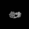
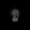
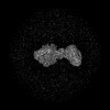
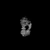
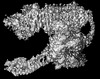
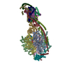
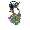
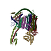
Journal: Cell Death Differ / Year: 2025 Title: Cryo-EM structure of the brine shrimp mitochondrial ATP synthase suggests an inactivation mechanism for the ATP synthase leak channel. Authors: Amrendra Kumar / Juliana da Fonseca Rezende E Mello / Yangyu Wu / Daniel Morris / Ikram Mezghani / Erin Smith / Stephane Rombauts / Peter Bossier / Juno Krahn / Fred J Sigworth / Nelli Mnatsakanyan / Abstract: Mammalian mitochondria undergo Ca-induced and cyclosporinA (CsA)-regulated permeability transition (mPT) by activating the mitochondrial permeability transition pore (mPTP) situated in mitochondrial ...Mammalian mitochondria undergo Ca-induced and cyclosporinA (CsA)-regulated permeability transition (mPT) by activating the mitochondrial permeability transition pore (mPTP) situated in mitochondrial inner membranes. Ca-induced prolonged openings of mPTP under certain pathological conditions result in mitochondrial swelling and rupture of the outer membrane, leading to mitochondrial dysfunction and cell death. While the exact molecular composition and structure of mPTP remain unknown, mammalian ATP synthase was reported to form voltage and Ca-activated leak channels involved in mPT. Unlike in mammals, mitochondria of the crustacean Artemia franciscana have the ability to accumulate large amounts of Ca without undergoing the mPT. Here, we performed structural and functional analysis of A. franciscana ATP synthase to study the molecular mechanism of mPTP inhibition in this organism. We found that the channel formed by the A. franciscana ATP synthase dwells predominantly in its inactive state and is insensitive to Ca, in contrast to porcine heart ATP synthase. Single-particle cryo-electron microscopy (cryo-EM) analysis revealed distinct structural features in A. franciscana ATP synthase compared with mammals. The stronger density of the e-subunit C-terminal region and its enhanced interaction with the c-ring were found in A. franciscana ATP synthase. These data suggest an inactivation mechanism of the ATP synthase leak channel and its possible contribution to the lack of mPT in this organism.
In the structure databanks used in Yorodumi, some data are registered as the other names, "COVID-19 virus" and "2019-nCoV". Here are the details of the virus and the list of structure data.
Jan 31, 2019. EMDB accession codes are about to change! (news from PDBe EMDB page)
EMDB accession codes are about to change! (news from PDBe EMDB page)
The allocation of 4 digits for EMDB accession codes will soon come to an end. Whilst these codes will remain in use, new EMDB accession codes will include an additional digit and will expand incrementally as the available range of codes is exhausted. The current 4-digit format prefixed with “EMD-” (i.e. EMD-XXXX) will advance to a 5-digit format (i.e. EMD-XXXXX), and so on. It is currently estimated that the 4-digit codes will be depleted around Spring 2019, at which point the 5-digit format will come into force.
The EM Navigator/Yorodumi systems omit the EMD- prefix.
Related info.:Q: What is EMD? / ID/Accession-code notation in Yorodumi/EM Navigator
Yorodumi is a browser for structure data from EMDB, PDB, SASBDB, etc.
This page is also the successor to EM Navigator detail page, and also detail information page/front-end page for Omokage search.
The word "yorodu" (or yorozu) is an old Japanese word meaning "ten thousand". "mi" (miru) is to see.
Related info.:EMDB / PDB / SASBDB / Comparison of 3 databanks / Yorodumi Search / Aug 31, 2016. New EM Navigator & Yorodumi / Yorodumi Papers / Jmol/JSmol / Function and homology information / Changes in new EM Navigator and Yorodumi
 Movie
Movie Controller


 Open data
 Basic information
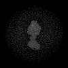
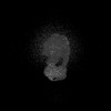
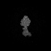
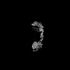
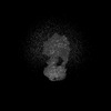
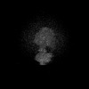
 Map data
 Sample
 Keywords
 Artemia franciscana (arthropod)
 Authors
 United States, 2 items
  Citation
 Structure visualization
 Downloads & links
 EMDB map data format
 emd_44170.png
 http://ftp.pdbj.org/pub/emdb/structures/EMD-44170
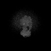

 Z (Sec.)
 Y (Row.)
 X (Col.)


 Sample components
 Processing
 Electron microscopy
 FIELD EMISSION GUN

