 Movie
Movie Controller


+ Open data

- Basic information
| Entry | Database: PDB / ID: 9az7 | ||||||
|---|---|---|---|---|---|---|---|
| Title | Chloride Sites in Photoactive Yellow Protein | ||||||
 Components Components | Photoactive yellow protein | ||||||
 Keywords Keywords | SIGNALING PROTEIN / Photoreceptor / Light Sensor / Chromophore / Photoreceptor Protein / Receptor / Sensory Transduction / PAS / LOV | ||||||
| Function / homology |  Function and homology information Function and homology informationphotoreceptor activity / phototransduction / regulation of DNA-templated transcription / identical protein binding Similarity search - Function | ||||||
| Biological species |  Halorhodospira halophila (bacteria) Halorhodospira halophila (bacteria) | ||||||
| Method |  X-RAY DIFFRACTION / MOLECULAR REPLACEMENT / Resolution: 2 Å X-RAY DIFFRACTION / MOLECULAR REPLACEMENT / Resolution: 2 Å | ||||||
 Authors Authors | Dyda, F. / Schotte, F. / Anfinrud, P. / Cho, H.S. | ||||||
| Funding support |  United States, 1items United States, 1items
| ||||||
 Citation Citation | Journal: Struct Dyn. / Year: 2024 Title: Watching a signaling protein function: What has been learned over four decades of time-resolved studies of photoactive yellow protein. Authors: Schotte, F. / Cho, H.S. / Dyda, F. / Anfinrud, P. | ||||||
| History |
|
- Structure visualization
| Structure viewer | Molecule: MolmilJmol/JSmol |
|---|
- Downloads & links
-Download
| PDBx/mmCIF format | 9az7.cif.gz | 50 KB | Display | PDBx/mmCIF format |
|---|---|---|---|---|
| PDB format | pdb9az7.ent.gz | 27.3 KB | Display | PDB format |
| PDBx/mmJSON format | 9az7.json.gz | Tree view | PDBx/mmJSON format | |
| Others |  Other downloads Other downloads |
-Validation report
| Arichive directory | https://data.pdbj.org/pub/pdb/validation_reports/az/9az7ftp://data.pdbj.org/pub/pdb/validation_reports/az/9az7 | HTTPS FTP |
|---|
-Related structure data

-Links
 PDBj
PDBj


- Assembly
| Deposited unit | 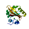
| ||||||||||||
|---|---|---|---|---|---|---|---|---|---|---|---|---|---|
| 1 |
| ||||||||||||
| Unit cell |
|
-Components
| #1: Protein | Mass: 13888.575 Da / Num. of mol.: 1 Source method: isolated from a genetically manipulated source Source: (gene. exp.) Halorhodospira halophila (bacteria) / Gene: pyp / Plasmid: PET16B / Production host: | ||||
|---|---|---|---|---|---|
| #2: Chemical | ChemComp-HC4 /   Mass: 164.158 Da / Num. of mol.: 1 / Source method: obtained synthetically / Formula: C9H8O3 Mass: 164.158 Da / Num. of mol.: 1 / Source method: obtained synthetically / Formula: C9H8O3 | ||||
| #3: Chemical |   Mass: 35.453 Da / Num. of mol.: 3 / Source method: obtained synthetically / Formula: Cl / Feature type: SUBJECT OF INVESTIGATION Mass: 35.453 Da / Num. of mol.: 3 / Source method: obtained synthetically / Formula: Cl / Feature type: SUBJECT OF INVESTIGATION#4: Water | ChemComp-HOH / |  Mass: 18.015 Da / Num. of mol.: 135 / Source method: isolated from a natural source / Formula: H2O Mass: 18.015 Da / Num. of mol.: 135 / Source method: isolated from a natural source / Formula: H2OHas ligand of interest | Y | |
-Experimental details
-Experiment
| Experiment | Method: X-RAY DIFFRACTION / Number of used crystals: 1 |
|---|
- Sample preparation
| Crystal | Density Matthews: 1.9 Å3/Da / Density % sol: 35.3 % |
|---|---|
| Crystal grow | Temperature: 293 K / Method: vapor diffusion, hanging drop / pH: 9 Details: 2.2 M ammonium sulfate, 1.0 M sodium chloride, 20 mM sodium phosphate |
-Data collection
| Diffraction | Mean temperature: 293 K / Serial crystal experiment: N |
|---|---|
| Diffraction source | Source: ROTATING ANODE / Type: RIGAKU FR-E SUPERBRIGHT / Wavelength: 1.54 Å |
| Detector | Type: RIGAKU SATURN A200 / Detector: CCD / Date: May 25, 2012 |
| Radiation | Monochromator: 1.54 / Protocol: SINGLE WAVELENGTH / Monochromatic (M) / Laue (L): M / Scattering type: x-ray |
| Radiation wavelength | Wavelength: 1.54 Å / Relative weight: 1 |
| Reflection | Resolution: 2→21.88 Å / Num. obs: 13007 / % possible obs: 94.4 % / Redundancy: 5.6 % / Biso Wilson estimate: 14.34 Å2 / CC1/2: 1 / Rmerge(I) obs: 0.026 / Net I/σ(I): 48.47 |
| Reflection shell | Resolution: 2→2.05 Å / Redundancy: 4.47 % / Rmerge(I) obs: 0.068 / Mean I/σ(I) obs: 20 / Num. unique obs: 702 / CC1/2: 0.995 / % possible all: 67.4 |
- Processing
| Software |
| ||||||||||||||||||||||||||||||||||||||||||||||||||||||||||||||||||||||
|---|---|---|---|---|---|---|---|---|---|---|---|---|---|---|---|---|---|---|---|---|---|---|---|---|---|---|---|---|---|---|---|---|---|---|---|---|---|---|---|---|---|---|---|---|---|---|---|---|---|---|---|---|---|---|---|---|---|---|---|---|---|---|---|---|---|---|---|---|---|---|---|
| Refinement | Method to determine structure: MOLECULAR REPLACEMENT / Resolution: 2→21.88 Å / SU ML: 0.1448 / Cross valid method: FREE R-VALUE / σ(F): 2.09 / Phase error: 18.3248 Stereochemistry target values: GeoStd + Monomer Library + CDL v1.2
| ||||||||||||||||||||||||||||||||||||||||||||||||||||||||||||||||||||||
| Solvent computation | Shrinkage radii: 0.9 Å / VDW probe radii: 1.1 Å / Solvent model: FLAT BULK SOLVENT MODEL | ||||||||||||||||||||||||||||||||||||||||||||||||||||||||||||||||||||||
| Displacement parameters | Biso mean: 20.08 Å2 | ||||||||||||||||||||||||||||||||||||||||||||||||||||||||||||||||||||||
| Refinement step | Cycle: LAST / Resolution: 2→21.88 Å
| ||||||||||||||||||||||||||||||||||||||||||||||||||||||||||||||||||||||
| Refine LS restraints |
| ||||||||||||||||||||||||||||||||||||||||||||||||||||||||||||||||||||||
| LS refinement shell |
|
