National Institutes of Health/National Heart, Lung, and Blood Institute (NIH/NHLBI)
米国
National Institutes of Health/National Institute of Arthritis and Musculoskeletal and Skin Diseases (NIH/NIAMS)
米国
引用
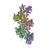
ジャーナル: Proc Natl Acad Sci U S A / 年: 2025 タイトル: Biochemical and structural bases for talin ABSs-F-actin interactions. 著者: Christian Biertümpfel / Yurika Yamada / Victor Vasquez-Montes / Thien Van Truong / A King Cada / Naoko Mizuno / 要旨: Focal adhesions (FAs) are large intracellular macromolecular assemblies that play a critical role in cell polarization and migration. Talin serves as a direct connection between integrin receptor and ...Focal adhesions (FAs) are large intracellular macromolecular assemblies that play a critical role in cell polarization and migration. Talin serves as a direct connection between integrin receptor and actomyosin cytoskeleton within FAs. Talin contains three actin-binding sites (ABS1-3) that engage discreetly during the development of FAs, thus acting as a critical player in FA initiation and maturation. However, the molecular basis of the ABS-F-actin interactions remains unknown. Here, we explore interactions of ABSs with F-actin to understand the multivalent behavior of talin. Particularly, the cryo-EM structure of the F-actin-ABS3 complex at 2.9 Å shows ABS3 spanning through two actin monomers along the filament axis, each occupied by the R13 rod subdomain and the DD domain. The dimerization of ABS3 occurs through the DD domain where both protomers interact on the actin surface, and the dimerization of talin to the actin surface is necessary for the engagement to F-actin. The R13 helical bundle is distorted upon binding to F-actin and releases the H1 helix from the rest of the bundle. This phenomenon has also been observed with other tension-sensing proteins like vinculin and α-catenin, highlighting that unfolding is relevant for its force sensing activity. On the contrary, ABS2 (R4R8 subdomains), which is thought to be critical for the maintenance of mature FAs, had multiple F-actin-binding regions within ABS2 and the binding likely occurred by these subdomains running through the surface of F-actin, thus strengthening the interactions upon the maturation of FAs.
濃度: 5 mg/ml / 包埋: NO / シャドウイング: NO / 染色: NO / 凍結: YES 詳細: G-actin was polymerized for 60 min at RT. 90 uM talin fragments were then incubated with 10 uM F-actin for 30 min at RT and ultra-centrifuged at 100,000 x g for 20 minutes at RT
 ムービー
ムービー コントローラー


 データを開く
 基本情報
基本情報 要素
要素 キーワード
キーワード 機能・相同性情報
 データ登録者
 米国, 2件
  引用
引用 構造の表示
構造の表示 ダウンロードとリンク
 その他のダウンロード
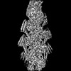


 PDBj
PDBj


 集合体
集合体


 分子量: 427.201 Da / 分子数: 5 / 由来タイプ: 合成 / 式: C10H15N5O10P2 / タイプ: SUBJECT OF INVESTIGATION / コメント: ADP, エネルギー貯蔵分子*YM
 分子量: 24.305 Da / 分子数: 5 / 由来タイプ: 合成 / 式: Mg / タイプ: SUBJECT OF INVESTIGATION
 試料調製
試料調製 電子顕微鏡撮影
 FIELD EMISSION GUN / 加速電圧: 300 kV / 照射モード: FLOOD BEAM
 解析
解析