National Institutes of Health/National Heart, Lung, and Blood Institute (NIH/NHLBI)
United States
National Institutes of Health/National Institute of Arthritis and Musculoskeletal and Skin Diseases (NIH/NIAMS)
United States
Citation
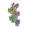
Journal: Proc Natl Acad Sci U S A / Year: 2025 Title: Biochemical and structural bases for talin ABSs-F-actin interactions. Authors: Christian Biertümpfel / Yurika Yamada / Victor Vasquez-Montes / Thien Van Truong / A King Cada / Naoko Mizuno / Abstract: Focal adhesions (FAs) are large intracellular macromolecular assemblies that play a critical role in cell polarization and migration. Talin serves as a direct connection between integrin receptor and ...Focal adhesions (FAs) are large intracellular macromolecular assemblies that play a critical role in cell polarization and migration. Talin serves as a direct connection between integrin receptor and actomyosin cytoskeleton within FAs. Talin contains three actin-binding sites (ABS1-3) that engage discreetly during the development of FAs, thus acting as a critical player in FA initiation and maturation. However, the molecular basis of the ABS-F-actin interactions remains unknown. Here, we explore interactions of ABSs with F-actin to understand the multivalent behavior of talin. Particularly, the cryo-EM structure of the F-actin-ABS3 complex at 2.9 Å shows ABS3 spanning through two actin monomers along the filament axis, each occupied by the R13 rod subdomain and the DD domain. The dimerization of ABS3 occurs through the DD domain where both protomers interact on the actin surface, and the dimerization of talin to the actin surface is necessary for the engagement to F-actin. The R13 helical bundle is distorted upon binding to F-actin and releases the H1 helix from the rest of the bundle. This phenomenon has also been observed with other tension-sensing proteins like vinculin and α-catenin, highlighting that unfolding is relevant for its force sensing activity. On the contrary, ABS2 (R4R8 subdomains), which is thought to be critical for the maintenance of mature FAs, had multiple F-actin-binding regions within ABS2 and the binding likely occurred by these subdomains running through the surface of F-actin, thus strengthening the interactions upon the maturation of FAs.
Conc.: 5 mg/ml / Embedding applied: NO / Shadowing applied: NO / Staining applied: NO / Vitrification applied: YES Details: G-actin was polymerized for 60 min at RT. 90 uM talin fragments were then incubated with 10 uM F-actin for 30 min at RT and ultra-centrifuged at 100,000 x g for 20 minutes at RT
Instrument: FEI VITROBOT MARK IV / Cryogen name: ETHANE / Humidity: 90 % / Chamber temperature: 277 K Details: 3.0 ul sample, blotted for 5 s from both sides with filter paper Whatman No.1
-
Electron microscopy imaging
Experimental equipment
Model: Titan Krios / Image courtesy: FEI Company
Microscopy
Model: FEI TITAN KRIOS
Electron gun
Electron source: FIELD EMISSION GUN / Accelerating voltage: 300 kV / Illumination mode: FLOOD BEAM
Electron lens
Mode: BRIGHT FIELD / Nominal magnification: 105000 X / Nominal defocus max: 2600 nm / Nominal defocus min: 800 nm / Alignment procedure: COMA FREE
Average exposure time: 6.6 sec. / Electron dose: 52 e/Å2 / Film or detector model: GATAN K3 BIOCONTINUUM (6k x 4k) / Num. of grids imaged: 1 / Num. of real images: 6767
EM imaging optics
Energyfilter name: GIF Bioquantum
-
Processing
EM software
ID
Name
Version
Category
1
RELION
3.1.0
particleselection
2
SerialEM
imageacquisition
4
CTFFIND
4
CTFcorrection
7
Coot
0.9.8.6
modelfitting
9
RELION
3.1.0
initialEulerassignment
10
RELION
3.1.0
finalEulerassignment
11
RELION
3.1.0
classification
12
RELION
3.1.0
3Dreconstruction
13
PHENIX
1.21-5207
modelrefinement
CTF correction
Type: PHASE FLIPPING AND AMPLITUDE CORRECTION
Helical symmerty
ID
Image processing-ID
Angular rotation/subunit (°)
Axial rise/subunit (Å)
Axial symmetry
1
1
-166.66
27.6
C1
2
1
-166.66
27.6
C1
Particle selection
Num. of particles selected: 2280210
3D reconstruction
Resolution: 2.98 Å / Resolution method: FSC 0.143 CUT-OFF / Num. of particles: 206326 / Algorithm: BACK PROJECTION / Num. of class averages: 1 / Symmetry type: HELICAL
In the structure databanks used in Yorodumi, some data are registered as the other names, "COVID-19 virus" and "2019-nCoV". Here are the details of the virus and the list of structure data.
Jan 31, 2019. EMDB accession codes are about to change! (news from PDBe EMDB page)
EMDB accession codes are about to change! (news from PDBe EMDB page)
The allocation of 4 digits for EMDB accession codes will soon come to an end. Whilst these codes will remain in use, new EMDB accession codes will include an additional digit and will expand incrementally as the available range of codes is exhausted. The current 4-digit format prefixed with “EMD-” (i.e. EMD-XXXX) will advance to a 5-digit format (i.e. EMD-XXXXX), and so on. It is currently estimated that the 4-digit codes will be depleted around Spring 2019, at which point the 5-digit format will come into force.
The EM Navigator/Yorodumi systems omit the EMD- prefix.
Related info.:Q: What is EMD? / ID/Accession-code notation in Yorodumi/EM Navigator
Yorodumi is a browser for structure data from EMDB, PDB, SASBDB, etc.
This page is also the successor to EM Navigator detail page, and also detail information page/front-end page for Omokage search.
The word "yorodu" (or yorozu) is an old Japanese word meaning "ten thousand". "mi" (miru) is to see.
Related info.:EMDB / PDB / SASBDB / Comparison of 3 databanks / Yorodumi Search / Aug 31, 2016. New EM Navigator & Yorodumi / Yorodumi Papers / Jmol/JSmol / Function and homology information / Changes in new EM Navigator and Yorodumi
 Movie
Movie Controller


 Open data
 Basic information
 Components
 Keywords
 Function and homology information
 Authors
 United States, 2items
  Citation
 Structure visualization
 Downloads & links
 Other downloads
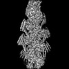


 PDBj
PDBj


 Assembly


 Mass: 427.201 Da / Num. of mol.: 5 / Source method: obtained synthetically / Formula: C10H15N5O10P2 / Feature type: SUBJECT OF INVESTIGATION / Comment: ADP, energy-carrying molecule*YM
 Mass: 24.305 Da / Num. of mol.: 5 / Source method: obtained synthetically / Formula: Mg / Feature type: SUBJECT OF INVESTIGATION
 Sample preparation
 Electron microscopy imaging
 FIELD EMISSION GUN / Accelerating voltage: 300 kV / Illumination mode: FLOOD BEAM
 Processing