 Movie
Movie Controller


+ Open data

- Basic information
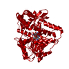
| Entry | Database: PDB / ID: 9aue | ||||||||||||||||||||||||||||||
|---|---|---|---|---|---|---|---|---|---|---|---|---|---|---|---|---|---|---|---|---|---|---|---|---|---|---|---|---|---|---|---|
| Title | Crystal structure of the holo form of GenB2 in complex with PMP | ||||||||||||||||||||||||||||||
 Components Components | 6'-epimerase, C-6' aminotransferase | ||||||||||||||||||||||||||||||
 Keywords Keywords | ANTIBIOTIC / gentamicin biosynthesis / holo form / Pyridoxamine-5'-phosphate (PMP) | ||||||||||||||||||||||||||||||
| Function / homology | Aminotransferase class-III / Aminotransferase class-III / transaminase activity / Pyridoxal phosphate-dependent transferase, small domain / Pyridoxal phosphate-dependent transferase, major domain / Pyridoxal phosphate-dependent transferase / pyridoxal phosphate binding / 4'-DEOXY-4'-AMINOPYRIDOXAL-5'-PHOSPHATE / 6'-epimerase, C-6' aminotransferase Function and homology information Function and homology information | ||||||||||||||||||||||||||||||
| Biological species |  Micromonospora echinospora (bacteria) Micromonospora echinospora (bacteria) | ||||||||||||||||||||||||||||||
| Method |  X-RAY DIFFRACTION / SYNCHROTRON / MOLECULAR REPLACEMENT / Resolution: 1.46 Å X-RAY DIFFRACTION / SYNCHROTRON / MOLECULAR REPLACEMENT / Resolution: 1.46 Å | ||||||||||||||||||||||||||||||
 Authors Authors | Oliveira, G.S. / Bury, P.S. / Huang, F. / Li, Y. / Araujo, N.C. / Zhou, J. / Sun, Y. / Leeper, F. / Leadlay, P. / Dias, M.V.B. | ||||||||||||||||||||||||||||||
| Funding support |  Brazil, Brazil,  China, 9items China, 9items
| ||||||||||||||||||||||||||||||
 Citation Citation | Journal: Acs Chem.Biol. / Year: 2024 Title: Structural and Functional Basis of GenB2 Isomerase Activity from Gentamicin Biosynthesis. Authors: Oliveira, G.S. / Dos S Bury, P. / Huang, F. / Li, Y. / Araujo, N.C. / Zhou, J. / Sun, Y. / Leeper, F.J. / Leadlay, P.F. / Dias, M.V.B. | ||||||||||||||||||||||||||||||
| History |
|
- Structure visualization
| Structure viewer | Molecule: MolmilJmol/JSmol |
|---|
- Downloads & links
-Download
| PDBx/mmCIF format | 9aue.cif.gz | 183.2 KB | Display | PDBx/mmCIF format |
|---|---|---|---|---|
| PDB format | pdb9aue.ent.gz | 142.7 KB | Display | PDB format |
| PDBx/mmJSON format | 9aue.json.gz | Tree view | PDBx/mmJSON format | |
| Others |  Other downloads Other downloads |
-Validation report
| Arichive directory | https://data.pdbj.org/pub/pdb/validation_reports/au/9aueftp://data.pdbj.org/pub/pdb/validation_reports/au/9aue | HTTPS FTP |
|---|
-Related structure data

-Links
 PDBj
PDBj- Assembly
| Deposited unit | 
| ||||||||||||
|---|---|---|---|---|---|---|---|---|---|---|---|---|---|
| 1 |
| ||||||||||||
| Unit cell |
| ||||||||||||
| Components on special symmetry positions |
|
-Components
| #1: Protein | Mass: 44701.285 Da / Num. of mol.: 1 Source method: isolated from a genetically manipulated source Source: (gene. exp.) Micromonospora echinospora (bacteria) / Gene: gacE, genB2, gntL / Production host: | ||||
|---|---|---|---|---|---|
| #2: Chemical | ChemComp-GOL /   Mass: 92.094 Da / Num. of mol.: 1 / Source method: obtained synthetically / Formula: C3H8O3 Mass: 92.094 Da / Num. of mol.: 1 / Source method: obtained synthetically / Formula: C3H8O3 | ||||
| #3: Chemical | ChemComp-PMP /   Mass: 248.173 Da / Num. of mol.: 1 / Source method: obtained synthetically / Formula: C8H13N2O5P / Feature type: SUBJECT OF INVESTIGATION Mass: 248.173 Da / Num. of mol.: 1 / Source method: obtained synthetically / Formula: C8H13N2O5P / Feature type: SUBJECT OF INVESTIGATION | ||||
| #4: Chemical |   Mass: 35.453 Da / Num. of mol.: 3 / Source method: obtained synthetically / Formula: Cl Mass: 35.453 Da / Num. of mol.: 3 / Source method: obtained synthetically / Formula: Cl#5: Water | ChemComp-HOH / |  Mass: 18.015 Da / Num. of mol.: 499 / Source method: isolated from a natural source / Formula: H2O Mass: 18.015 Da / Num. of mol.: 499 / Source method: isolated from a natural source / Formula: H2OHas ligand of interest | Y | |
-Experimental details
-Experiment
| Experiment | Method: X-RAY DIFFRACTION / Number of used crystals: 1 |
|---|
- Sample preparation
| Crystal | Density Matthews: 2.28 Å3/Da / Density % sol: 45.97 % |
|---|---|
| Crystal grow | Temperature: 291 K / Method: vapor diffusion, hanging drop Details: 0.1M PIPES, pH 6.0, 1 M NaCl, 29% PEG 4000 and 30% 6-aminohexanoic acid. |
-Data collection
| Diffraction | Mean temperature: 100 K / Serial crystal experiment: N |
|---|---|
| Diffraction source | Source: SYNCHROTRON / Site: LNLS SIRIUS / Beamline: MANACA / Wavelength: 0.97718 Å |
| Detector | Type: DECTRIS PILATUS 2M / Detector: PIXEL / Date: May 26, 2022 |
| Radiation | Protocol: SINGLE WAVELENGTH / Monochromatic (M) / Laue (L): M / Scattering type: x-ray |
| Radiation wavelength | Wavelength: 0.97718 Å / Relative weight: 1 |
| Reflection | Resolution: 1.45→46.43 Å / Num. obs: 71076 / % possible obs: 99 % / Redundancy: 13.4 % / CC1/2: 0.999 / Rmerge(I) obs: 0.145 / Rpim(I) all: 0.041 / Rrim(I) all: 0.151 / Χ2: 1 / Net I/σ(I): 10.8 |
| Reflection shell | Resolution: 1.45→1.47 Å / % possible obs: 94.5 % / Redundancy: 12.6 % / Rmerge(I) obs: 2.963 / Num. measured all: 43225 / Num. unique obs: 3429 / CC1/2: 0.41 / Rpim(I) all: 0.843 / Rrim(I) all: 3.085 / Χ2: 0.99 / Net I/σ(I) obs: 1 |
- Processing
| Software |
| |||||||||||||||||||||||||||||||||||||||||||||||||||||||||||||||||||||||||||||||||||||||||||||||||||||||||
|---|---|---|---|---|---|---|---|---|---|---|---|---|---|---|---|---|---|---|---|---|---|---|---|---|---|---|---|---|---|---|---|---|---|---|---|---|---|---|---|---|---|---|---|---|---|---|---|---|---|---|---|---|---|---|---|---|---|---|---|---|---|---|---|---|---|---|---|---|---|---|---|---|---|---|---|---|---|---|---|---|---|---|---|---|---|---|---|---|---|---|---|---|---|---|---|---|---|---|---|---|---|---|---|---|---|---|
| Refinement | Method to determine structure: MOLECULAR REPLACEMENT / Resolution: 1.46→46.43 Å / SU ML: 0.19 / Cross valid method: NONE / σ(F): 1.33 / Phase error: 19.88 / Stereochemistry target values: ML
| |||||||||||||||||||||||||||||||||||||||||||||||||||||||||||||||||||||||||||||||||||||||||||||||||||||||||
| Solvent computation | Shrinkage radii: 0.9 Å / VDW probe radii: 1.1 Å / Solvent model: FLAT BULK SOLVENT MODEL | |||||||||||||||||||||||||||||||||||||||||||||||||||||||||||||||||||||||||||||||||||||||||||||||||||||||||
| Refinement step | Cycle: LAST / Resolution: 1.46→46.43 Å
| |||||||||||||||||||||||||||||||||||||||||||||||||||||||||||||||||||||||||||||||||||||||||||||||||||||||||
| Refine LS restraints |
| |||||||||||||||||||||||||||||||||||||||||||||||||||||||||||||||||||||||||||||||||||||||||||||||||||||||||
| LS refinement shell |
|
