 Movie
Movie Controller

[English] 日本語
 Yorodumi
- PDB-9au9: Ternary complex of human DNA polymerase theta polymerase domain w... -
+ Open data

- Basic information
| Entry | Database: PDB / ID: 9au9 | |||||||||||||||||||||
|---|---|---|---|---|---|---|---|---|---|---|---|---|---|---|---|---|---|---|---|---|---|---|
| Title | Ternary complex of human DNA polymerase theta polymerase domain with a mismatched T:G base pair | |||||||||||||||||||||
 Components Components |
| |||||||||||||||||||||
 Keywords Keywords | DNA BINDING PROTEIN/DNA / DNA translesion synthesis / theta-mediated end joining / A-family DNA polymerase / DNA BINDING PROTEIN-DNA complex | |||||||||||||||||||||
| Function / homology |  Function and homology information Function and homology informationdouble-strand break repair via alternative nonhomologous end joining / HDR through MMEJ (alt-NHEJ) / single-stranded DNA helicase activity / replication fork processing / mitochondrial nucleoid / site of DNA damage / 5'-deoxyribose-5-phosphate lyase activity / error-prone translesion synthesis / negative regulation of double-strand break repair via homologous recombination / somatic hypermutation of immunoglobulin genes ...double-strand break repair via alternative nonhomologous end joining / HDR through MMEJ (alt-NHEJ) / single-stranded DNA helicase activity / replication fork processing / mitochondrial nucleoid / site of DNA damage / 5'-deoxyribose-5-phosphate lyase activity / error-prone translesion synthesis / negative regulation of double-strand break repair via homologous recombination / somatic hypermutation of immunoglobulin genes / DNA helicase activity / base-excision repair / protein homooligomerization / RNA-directed DNA polymerase / RNA-directed DNA polymerase activity / double-strand break repair / site of double-strand break / DNA helicase / DNA-directed DNA polymerase / damaged DNA binding / DNA-directed DNA polymerase activity / DNA repair / DNA damage response / chromatin binding / magnesium ion binding / Golgi apparatus / ATP hydrolysis activity / nucleoplasm / ATP binding / identical protein binding / nucleus / cytosol Similarity search - Function | |||||||||||||||||||||
| Biological species |  Homo sapiens (human) Homo sapiens (human) | |||||||||||||||||||||
| Method | ELECTRON MICROSCOPY / single particle reconstruction / cryo EM / Resolution: 3.32 Å | |||||||||||||||||||||
 Authors Authors | Li, C. / Gao, Y. | |||||||||||||||||||||
| Funding support |  United States, 2items United States, 2items
| |||||||||||||||||||||
 Citation Citation | Journal: Nat Commun / Year: 2025 Title: Structural basis of error-prone DNA synthesis by DNA polymerase θ. Authors: Chuxuan Li / Leora M Maksoud / Yang Gao / Abstract: DNA polymerase θ (Pol θ) is an A-family DNA polymerase specialized in DNA double-strand breaks repair and translesion synthesis. Distinct from its high-fidelity homologs in DNA replication, Pol θ ...DNA polymerase θ (Pol θ) is an A-family DNA polymerase specialized in DNA double-strand breaks repair and translesion synthesis. Distinct from its high-fidelity homologs in DNA replication, Pol θ catalyzes template-dependent DNA synthesis with an inherent propensity for error incorporation. However, the structural basis of Pol θ's low-fidelity DNA synthesis is not clear. Here, we present cryo-electron microscopy structures detailing the polymerase domain of human Pol θ in complex with a cognate C:G base pair (bp), a mismatched T:G bp, or a mismatched T:T bp. Our structures illustrate that Pol θ snugly accommodates the mismatched nascent base pairs within its active site with the finger domain well-closed, consistent with our in-solution fluorescence measurement but in contrast to its high-fidelity homologs. In addition, structural examination and mutagenesis study show that unique residues surrounding the active site contribute to the stabilization of the mismatched nascent base pair. Furthermore, Pol θ can efficiently extend from the misincorporated T:G or T:T mismatches, yet with a preference for template or primer looping-out, resulting in insertions and deletions. Collectively, our results elucidate how an A-family polymerase is adapted for error-prone DNA synthesis. | |||||||||||||||||||||
| History |
|
- Structure visualization
| Structure viewer | Molecule: MolmilJmol/JSmol |
|---|
- Downloads & links
-Download
| PDBx/mmCIF format | 9au9.cif.gz | 165.1 KB | Display | PDBx/mmCIF format |
|---|---|---|---|---|
| PDB format | pdb9au9.ent.gz | 119.9 KB | Display | PDB format |
| PDBx/mmJSON format | 9au9.json.gz | Tree view | PDBx/mmJSON format | |
| Others |  Other downloads Other downloads |
-Validation report
| Arichive directory | https://data.pdbj.org/pub/pdb/validation_reports/au/9au9ftp://data.pdbj.org/pub/pdb/validation_reports/au/9au9 | HTTPS FTP |
|---|
-Related structure data
| Related structure data |  43875MC  9au5C  9au8C M: map data used to model this data C: citing same article ( |
|---|---|
| Similar structure data |


-Links
 PDBj
PDBj


- Assembly
| Deposited unit | 
|
|---|---|
| 1 |
|
-Components
| #1: Protein | Mass: 89585.555 Da / Num. of mol.: 1 Source method: isolated from a genetically manipulated source Source: (gene. exp.) Homo sapiens (human) / Gene: POLQ, POLHProduction host:  References: UniProt: O75417, DNA-directed DNA polymerase |
|---|---|
| #2: DNA chain | Mass: 6149.978 Da / Num. of mol.: 1 / Source method: obtained synthetically / Source: (synth.) Homo sapiens (human) |
| #3: DNA chain | Mass: 8904.736 Da / Num. of mol.: 1 / Source method: obtained synthetically / Source: (synth.) Homo sapiens (human) |
| #4: Chemical | ChemComp-DGT / 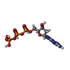  Mass: 507.181 Da / Num. of mol.: 1 / Source method: obtained synthetically / Formula: C10H16N5O13P3 / Feature type: SUBJECT OF INVESTIGATION Mass: 507.181 Da / Num. of mol.: 1 / Source method: obtained synthetically / Formula: C10H16N5O13P3 / Feature type: SUBJECT OF INVESTIGATION |
| Has ligand of interest | Y |
| Has protein modification | N |
-Experimental details
-Experiment
| Experiment | Method: ELECTRON MICROSCOPY |
|---|---|
| EM experiment | Aggregation state: PARTICLE / 3D reconstruction method: single particle reconstruction |
- Sample preparation
| Component | Name: Ternary complex of human DNA polymerase theta with duplex DNA and incoming nucleotide Type: COMPLEX / Entity ID: #1-#3 / Source: RECOMBINANT |
|---|---|
| Source (natural) | Organism: Homo sapiens (human) |
| Source (recombinant) | Organism: |
| Buffer solution | pH: 7.6 |
| Specimen | Conc.: 0.7 mg/ml / Embedding applied: NO / Shadowing applied: NO / Staining applied: NO / Vitrification applied: YES |
| Vitrification | Cryogen name: ETHANE / Humidity: 100 % / Chamber temperature: 295 K |
- Electron microscopy imaging
| Experimental equipment |  Model: Titan Krios / Image courtesy: FEI Company |
|---|---|
| Microscopy | Model: TFS KRIOS |
| Electron gun | Electron source:  FIELD EMISSION GUN / Accelerating voltage: 300 kV / Illumination mode: FLOOD BEAM FIELD EMISSION GUN / Accelerating voltage: 300 kV / Illumination mode: FLOOD BEAM |
| Electron lens | Mode: BRIGHT FIELD / Nominal defocus max: 2000 nm / Nominal defocus min: 800 nm |
| Image recording | Electron dose: 1.2 e/Å2 / Film or detector model: GATAN K3 (6k x 4k) |
- Processing
| EM software | Name: PHENIX / Version: 1.21_5207: / Category: model refinement |
|---|---|
| CTF correction | Type: PHASE FLIPPING AND AMPLITUDE CORRECTION |
| 3D reconstruction | Resolution: 3.32 Å / Resolution method: FSC 0.143 CUT-OFF / Num. of particles: 93767 / Symmetry type: POINT |
