 Movie
Movie Controller


+ Open data

- Basic information
| Entry | Database: PDB / ID: 8zxm | |||||||||||||||||||||
|---|---|---|---|---|---|---|---|---|---|---|---|---|---|---|---|---|---|---|---|---|---|---|
| Title | Cryo-EM structure of human GLUT9 bound to urate | |||||||||||||||||||||
 Components Components | Solute carrier family 2, facilitated glucose transporter member 9 | |||||||||||||||||||||
 Keywords Keywords | TRANSPORT PROTEIN / Urate / Transporter / Hypouricemia / RHUC | |||||||||||||||||||||
| Function / homology |  Function and homology information Function and homology informationDefective SLC2A9 causes hypouricemia renal 2 (RHUC2) / fructose transmembrane transporter activity / fructose transmembrane transport / dehydroascorbic acid transport / hexose transmembrane transport / carbohydrate:proton symporter activity / Cellular hexose transport / D-glucose transmembrane transporter activity / D-glucose transmembrane transport / urate transport ...Defective SLC2A9 causes hypouricemia renal 2 (RHUC2) / fructose transmembrane transporter activity / fructose transmembrane transport / dehydroascorbic acid transport / hexose transmembrane transport / carbohydrate:proton symporter activity / Cellular hexose transport / D-glucose transmembrane transporter activity / D-glucose transmembrane transport / urate transport / urate metabolic process / urate transmembrane transporter activity / : / transmembrane transporter activity / basolateral plasma membrane / apical plasma membrane / membrane / plasma membrane Similarity search - Function | |||||||||||||||||||||
| Biological species |  Homo sapiens (human) Homo sapiens (human) | |||||||||||||||||||||
| Method | ELECTRON MICROSCOPY / single particle reconstruction / cryo EM / Resolution: 3.39 Å | |||||||||||||||||||||
 Authors Authors | Matsushita, D. / Lee, Y. / Nishizawa, T. | |||||||||||||||||||||
| Funding support |  Japan, 6items Japan, 6items
| |||||||||||||||||||||
 Citation Citation | Journal: Cell Rep / Year: 2025 Title: Structural basis of urate transport by glucose transporter 9. Authors: Daiki Matsushita / Yu Toyoda / Yongchan Lee / Maeda Aoi / Hirotaka Matsuo / Tappei Takada / Tomohiro Nishizawa / Abstract: Glucose transporter 9 (GLUT9) is a critical urate transporter involved in renal reabsorption, playing a pivotal role in regulating physiological urate levels and representing a potential therapeutic ...Glucose transporter 9 (GLUT9) is a critical urate transporter involved in renal reabsorption, playing a pivotal role in regulating physiological urate levels and representing a potential therapeutic target for gout. Despite such clinical significance, the structural basis of urate recognition and transport by GLUT9 remains elusive. Here, we present the cryoelectron microscopy (cryo-EM) structures of GLUT9 in the inward-open conformation in both apo and urate-bound states. Urate binds in a cleft between the N-terminal and C-terminal domains, interacting via hydrogen bonds and hydrophobic interactions. Structural comparison with sugar-transporting GLUTs highlights unique amino acid compositions in the substrate recognition pocket of GLUT9. Functional and mutational studies directly measuring GLUT9-mediated urate uptake further demonstrate the cooperative roles of multiple residues in urate recognition. Our findings elucidate the structural basis of urate transport by GLUT9 and provide valuable insights for the development of uricosuric drugs targeting GLUT9. | |||||||||||||||||||||
| History |
|
- Structure visualization
| Structure viewer | Molecule: MolmilJmol/JSmol |
|---|
- Downloads & links
-Download
| PDBx/mmCIF format | 8zxm.cif.gz | 107 KB | Display | PDBx/mmCIF format |
|---|---|---|---|---|
| PDB format | pdb8zxm.ent.gz | 78 KB | Display | PDB format |
| PDBx/mmJSON format | 8zxm.json.gz | Tree view | PDBx/mmJSON format | |
| Others |  Other downloads Other downloads |
-Validation report
| Arichive directory | https://data.pdbj.org/pub/pdb/validation_reports/zx/8zxmftp://data.pdbj.org/pub/pdb/validation_reports/zx/8zxm | HTTPS FTP |
|---|
-Related structure data
| Related structure data |  60544MC 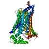 8zxnC M: map data used to model this data C: citing same article ( |
|---|---|
| Similar structure data |
-Links
 PDBj
PDBj


- Assembly
| Deposited unit | 
|
|---|---|
| 1 |
|
-Components
| #1: Protein | Mass: 59707.477 Da / Num. of mol.: 1 Source method: isolated from a genetically manipulated source Source: (gene. exp.) Homo sapiens (human) / Gene: SLC2A9, GLUT9 / Cell line (production host): HEK293F / Production host: Homo sapiens (human) / References: UniProt: Q9NRM0 |
|---|---|
| #2: Polysaccharide | 2-acetamido-2-deoxy-beta-D-glucopyranose-(1-4)-2-acetamido-2-deoxy-beta-D-glucopyranose Source method: isolated from a genetically manipulated source |
| #3: Chemical | ChemComp-URC /   Mass: 168.110 Da / Num. of mol.: 1 / Source method: obtained synthetically / Formula: C5H4N4O3 / Feature type: SUBJECT OF INVESTIGATION Mass: 168.110 Da / Num. of mol.: 1 / Source method: obtained synthetically / Formula: C5H4N4O3 / Feature type: SUBJECT OF INVESTIGATION |
| Has ligand of interest | Y |
| Has protein modification | Y |
-Experimental details
-Experiment
| Experiment | Method: ELECTRON MICROSCOPY |
|---|---|
| EM experiment | Aggregation state: PARTICLE / 3D reconstruction method: single particle reconstruction |
- Sample preparation
| Component | Name: Glucose transporter 9 / Type: COMPLEX / Entity ID: #1 / Source: RECOMBINANT |
|---|---|
| Molecular weight | Value: 60 kDa/nm / Experimental value: NO |
| Source (natural) | Organism: Homo sapiens (human) |
| Source (recombinant) | Organism: Homo sapiens (human) |
| Buffer solution | pH: 8 |
| Specimen | Embedding applied: NO / Shadowing applied: NO / Staining applied: NO / Vitrification applied: YES |
| Vitrification | Cryogen name: ETHANE |
- Electron microscopy imaging
| Experimental equipment |  Model: Titan Krios / Image courtesy: FEI Company |
|---|---|
| Microscopy | Model: FEI TITAN KRIOS |
| Electron gun | Electron source:  FIELD EMISSION GUN / Accelerating voltage: 300 kV / Illumination mode: FLOOD BEAM FIELD EMISSION GUN / Accelerating voltage: 300 kV / Illumination mode: FLOOD BEAM |
| Electron lens | Mode: BRIGHT FIELD / Nominal defocus max: 1600 nm / Nominal defocus min: 800 nm |
| Image recording | Electron dose: 51 e/Å2 / Film or detector model: GATAN K3 BIOQUANTUM (6k x 4k) |
- Processing
| CTF correction | Type: PHASE FLIPPING AND AMPLITUDE CORRECTION | ||||||||||||||||||||||||||||||||||||||||||||||||||||||||||||||||||||||||||||||||||||||||||||||||||||||||||
|---|---|---|---|---|---|---|---|---|---|---|---|---|---|---|---|---|---|---|---|---|---|---|---|---|---|---|---|---|---|---|---|---|---|---|---|---|---|---|---|---|---|---|---|---|---|---|---|---|---|---|---|---|---|---|---|---|---|---|---|---|---|---|---|---|---|---|---|---|---|---|---|---|---|---|---|---|---|---|---|---|---|---|---|---|---|---|---|---|---|---|---|---|---|---|---|---|---|---|---|---|---|---|---|---|---|---|---|
| 3D reconstruction | Resolution: 3.39 Å / Resolution method: FSC 0.143 CUT-OFF / Num. of particles: 180531 / Symmetry type: POINT | ||||||||||||||||||||||||||||||||||||||||||||||||||||||||||||||||||||||||||||||||||||||||||||||||||||||||||
| Refinement | Resolution: 3.39→3.39 Å / Cor.coef. Fo:Fc: 0.79 / SU B: 16.093 / SU ML: 0.253 / ESU R: 0.695 Stereochemistry target values: MAXIMUM LIKELIHOOD WITH PHASES Details: HYDROGENS HAVE BEEN USED IF PRESENT IN THE INPUT
| ||||||||||||||||||||||||||||||||||||||||||||||||||||||||||||||||||||||||||||||||||||||||||||||||||||||||||
| Solvent computation | Solvent model: PARAMETERS FOR MASK CACLULATION | ||||||||||||||||||||||||||||||||||||||||||||||||||||||||||||||||||||||||||||||||||||||||||||||||||||||||||
| Displacement parameters | Biso mean: 127.209 Å2 | ||||||||||||||||||||||||||||||||||||||||||||||||||||||||||||||||||||||||||||||||||||||||||||||||||||||||||
| Refinement step | Cycle: 1 / Total: 3666 | ||||||||||||||||||||||||||||||||||||||||||||||||||||||||||||||||||||||||||||||||||||||||||||||||||||||||||
| Refine LS restraints |
|