+ Open data
- Basic information
| Entry | Database: PDB / ID: 8zto | |||||||||||||||||||||
|---|---|---|---|---|---|---|---|---|---|---|---|---|---|---|---|---|---|---|---|---|---|---|
| Title | Cryo-EM structure of the human XPR1 | |||||||||||||||||||||
 Components Components | Solute carrier family 53 member 1 | |||||||||||||||||||||
 Keywords Keywords | STRUCTURAL PROTEIN / SLC transporter / XPR1 / SLC53A1 / Pi / exporter | |||||||||||||||||||||
| Function / homology |  Function and homology information Function and homology informationphosphate transmembrane transporter activity / phosphate ion transport / intracellular phosphate ion homeostasis / cellular response to phosphate starvation / phosphate ion transmembrane transport / inositol hexakisphosphate binding / efflux transmembrane transporter activity / response to virus / virus receptor activity / plasma membrane Similarity search - Function | |||||||||||||||||||||
| Biological species |  Homo sapiens (human) Homo sapiens (human) | |||||||||||||||||||||
| Method | ELECTRON MICROSCOPY / single particle reconstruction / cryo EM / Resolution: 3.55 Å | |||||||||||||||||||||
 Authors Authors | She, J. / Chen, L. | |||||||||||||||||||||
| Funding support |  China, 1items China, 1items
| |||||||||||||||||||||
 Citation Citation |  Journal: Nat Commun / Year: 2025 Journal: Nat Commun / Year: 2025Title: Structure and function of human XPR1 in phosphate export. Authors: Long Chen / Jin He / Mingxing Wang / Ji She /  Abstract: Xenotropic and polytropic retrovirus receptor 1 (XPR1) functions as a phosphate exporter and is pivotal in maintaining human phosphate homeostasis. It has been identified as a causative gene for ...Xenotropic and polytropic retrovirus receptor 1 (XPR1) functions as a phosphate exporter and is pivotal in maintaining human phosphate homeostasis. It has been identified as a causative gene for primary familial brain calcification. Here we present the cryogenic electron microscopy (cryo-EM) structure of human XPR1 (HsXPR1). HsXPR1 exhibits a dimeric structure in which only TM1 directly constitutes the dimer interface of the transmembrane domain. Each HsXPR1 subunit can be divided spatially into a core domain and a scaffold domain. The core domain of HsXPR1 forms a pore-like structure, along which two phosphate-binding sites enriched with positively charged residues are identified. Mutations of key residues at either site substantially diminish the transport activity of HsXPR1. Phosphate binding at the central site may trigger a conformational change at TM9, leading to the opening of the extracellular gate. In addition, our structural analysis reveals a new conformational state of HsXPR1 in which the cytoplasmic SPX domains form a V-shaped structure. Altogether, our results elucidate the overall architecture of HsXPR1 and shed light on XPR1-mediated phosphate export. | |||||||||||||||||||||
| History |
|
- Structure visualization
| Structure viewer | Molecule:  Molmil Molmil Jmol/JSmol Jmol/JSmol |
|---|
- Downloads & links
- Download
| PDBx/mmCIF format |  8zto.cif.gz 8zto.cif.gz | 163.1 KB | Display |  PDBx/mmCIF format PDBx/mmCIF format |
|---|---|---|---|---|
| PDB format |  pdb8zto.ent.gz pdb8zto.ent.gz | 123.6 KB | Display |  PDB format PDB format |
| PDBx/mmJSON format |  8zto.json.gz 8zto.json.gz | Tree view |  PDBx/mmJSON format PDBx/mmJSON format | |
| Others |  Other downloads Other downloads |
-Validation report
| Arichive directory |  https://data.pdbj.org/pub/pdb/validation_reports/zt/8zto https://data.pdbj.org/pub/pdb/validation_reports/zt/8zto ftp://data.pdbj.org/pub/pdb/validation_reports/zt/8zto ftp://data.pdbj.org/pub/pdb/validation_reports/zt/8zto | HTTPS FTP |
|---|
-Related structure data
| Related structure data |  60469MC M: map data used to model this data C: citing same article ( |
|---|---|
| Similar structure data | Similarity search - Function & homology  F&H Search F&H Search |
- Links
Links
- Assembly
| Deposited unit | 
|
|---|---|
| 1 |
|
- Components
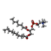
| #1: Protein | Mass: 82789.992 Da / Num. of mol.: 2 Source method: isolated from a genetically manipulated source Source: (gene. exp.)  Homo sapiens (human) / Gene: XPR1, SLC53A1, SYG1, X3 / Production host: Homo sapiens (human) / Gene: XPR1, SLC53A1, SYG1, X3 / Production host:  Homo sapiens (human) / References: UniProt: Q9UBH6 Homo sapiens (human) / References: UniProt: Q9UBH6#2: Chemical | #3: Chemical | Has ligand of interest | Y | Has protein modification | N | |
|---|
-Experimental details
-Experiment
| Experiment | Method: ELECTRON MICROSCOPY |
|---|---|
| EM experiment | Aggregation state: PARTICLE / 3D reconstruction method: single particle reconstruction |
- Sample preparation
| Component | Name: The structure of Human XPR1 / Type: COMPLEX / Entity ID: #1 / Source: RECOMBINANT |
|---|---|
| Source (natural) | Organism:  Homo sapiens (human) Homo sapiens (human) |
| Source (recombinant) | Organism:  Homo sapiens (human) Homo sapiens (human) |
| Buffer solution | pH: 8 |
| Specimen | Embedding applied: NO / Shadowing applied: NO / Staining applied: NO / Vitrification applied: YES |
| Vitrification | Cryogen name: ETHANE |
- Electron microscopy imaging
| Experimental equipment |  Model: Titan Krios / Image courtesy: FEI Company |
|---|---|
| Microscopy | Model: FEI TITAN KRIOS |
| Electron gun | Electron source:  FIELD EMISSION GUN / Accelerating voltage: 300 kV / Illumination mode: FLOOD BEAM FIELD EMISSION GUN / Accelerating voltage: 300 kV / Illumination mode: FLOOD BEAM |
| Electron lens | Mode: BRIGHT FIELD / Nominal defocus max: 2800 nm / Nominal defocus min: 1400 nm |
| Image recording | Electron dose: 55 e/Å2 / Film or detector model: GATAN K3 BIOQUANTUM (6k x 4k) |
- Processing
| EM software | Name: PHENIX / Category: model refinement | ||||||||||||||||||||||||
|---|---|---|---|---|---|---|---|---|---|---|---|---|---|---|---|---|---|---|---|---|---|---|---|---|---|
| CTF correction | Type: NONE | ||||||||||||||||||||||||
| 3D reconstruction | Resolution: 3.55 Å / Resolution method: FSC 0.143 CUT-OFF / Num. of particles: 424758 / Symmetry type: POINT | ||||||||||||||||||||||||
| Refine LS restraints |
|
 Movie
Movie Controller


 PDBj
PDBj


