 Movie
Movie Controller


+ Open data

- Basic information
| Entry | Database: PDB / ID: 8zpg | ||||||
|---|---|---|---|---|---|---|---|
| Title | SFX reaction state structure (20-40min) of alanine racemase | ||||||
 Components Components | Alanine racemase 2 | ||||||
 Keywords Keywords | ISOMERASE / enzyme / substrate complex / peptidodiglycan synthesis | ||||||
| Function / homology |  Function and homology information Function and homology informationalanine racemase / D-alanine biosynthetic process / alanine racemase activity / peptidoglycan biosynthetic process / pyridoxal phosphate binding / cytosol Similarity search - Function | ||||||
| Biological species |  | ||||||
| Method |  X-RAY DIFFRACTION / FREE ELECTRON LASER / MOLECULAR REPLACEMENT / Resolution: 2.3 Å X-RAY DIFFRACTION / FREE ELECTRON LASER / MOLECULAR REPLACEMENT / Resolution: 2.3 Å | ||||||
 Authors Authors | Kim, J. / Nam, K.H. / Cho, Y. | ||||||
| Funding support |  Korea, Republic Of, 1items Korea, Republic Of, 1items
| ||||||
 Citation Citation | Journal: Sci Rep / Year: 2024 Title: Exploring the reaction dynamics of alanine racemase using serial femtosecond crystallography. Authors: Kim, J. / Park, J. / Lee, K. / Chung, W.K. / Nam, K.H. / Cho, Y. | ||||||
| History |
|
- Structure visualization
| Structure viewer | Molecule: MolmilJmol/JSmol |
|---|
- Downloads & links
-Download
| PDBx/mmCIF format | 8zpg.cif.gz | 305.6 KB | Display | PDBx/mmCIF format |
|---|---|---|---|---|
| PDB format | pdb8zpg.ent.gz | 245.7 KB | Display | PDB format |
| PDBx/mmJSON format | 8zpg.json.gz | Tree view | PDBx/mmJSON format | |
| Others |  Other downloads Other downloads |
-Validation report
| Summary document | 8zpg_validation.pdf.gz | 2.4 MB | Display | wwPDB validaton report |
|---|---|---|---|---|
| Full document | 8zpg_full_validation.pdf.gz | 2.4 MB | Display | |
| Data in XML | 8zpg_validation.xml.gz | 61.6 KB | Display | |
| Data in CIF | 8zpg_validation.cif.gz | 79.9 KB | Display | |
| Arichive directory | https://data.pdbj.org/pub/pdb/validation_reports/zp/8zpgftp://data.pdbj.org/pub/pdb/validation_reports/zp/8zpg | HTTPS FTP |
-Related structure data
| Related structure data |  8zpeC  8zpfC 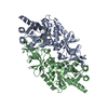 8zphC  9jt7C  6q72S C: citing same article ( S: Starting model for refinement |
|---|---|
| Similar structure data |
-Links
 PDBj
PDBj- Assembly
| Deposited unit | 
| ||||||||
|---|---|---|---|---|---|---|---|---|---|
| 1 | 
| ||||||||
| 2 | 
| ||||||||
| Unit cell |
|
-Components
| #1: Protein | Mass: 44122.312 Da / Num. of mol.: 4 Source method: isolated from a genetically manipulated source Source: (gene. exp.) Gene: alr2, yncD, BSU17640 / Production host: #2: Chemical | ChemComp-PLP /   Mass: 247.142 Da / Num. of mol.: 4 / Source method: obtained synthetically / Formula: C8H10NO6P / Feature type: SUBJECT OF INVESTIGATION Mass: 247.142 Da / Num. of mol.: 4 / Source method: obtained synthetically / Formula: C8H10NO6P / Feature type: SUBJECT OF INVESTIGATION#3: Chemical |   Type: L-peptide linking / Mass: 89.093 Da / Num. of mol.: 3 / Source method: obtained synthetically / Formula: C3H7NO2 / Feature type: SUBJECT OF INVESTIGATION Type: L-peptide linking / Mass: 89.093 Da / Num. of mol.: 3 / Source method: obtained synthetically / Formula: C3H7NO2 / Feature type: SUBJECT OF INVESTIGATION#4: Chemical | ChemComp-CL /   Mass: 35.453 Da / Num. of mol.: 4 / Source method: obtained synthetically / Formula: Cl Mass: 35.453 Da / Num. of mol.: 4 / Source method: obtained synthetically / Formula: Cl#5: Water | ChemComp-HOH / |  Mass: 18.015 Da / Num. of mol.: 78 / Source method: isolated from a natural source / Formula: H2O Mass: 18.015 Da / Num. of mol.: 78 / Source method: isolated from a natural source / Formula: H2OHas ligand of interest | Y | Has protein modification | Y | |
|---|
-Experimental details
-Experiment
| Experiment | Method: X-RAY DIFFRACTION / Number of used crystals: 1 |
|---|
- Sample preparation
| Crystal | Density Matthews: 2.44 Å3/Da / Density % sol: 49.64 % |
|---|---|
| Crystal grow | Temperature: 293 K / Method: batch mode / pH: 8.5 Details: 100 mM Tris-Cl (pH 8.5), 0.25 M MgCl2, and 25% Peg 4K |
-Data collection
| Diffraction | Mean temperature: 293 K / Serial crystal experiment: Y |
|---|---|
| Diffraction source | Source: FREE ELECTRON LASER / Site: PAL-XFEL / Beamline: NCI / Wavelength: 1 Å |
| Detector | Type: RAYONIX MX225-HS / Detector: CCD / Date: Apr 20, 2021 |
| Radiation | Protocol: SINGLE WAVELENGTH / Monochromatic (M) / Laue (L): M / Scattering type: x-ray |
| Radiation wavelength | Wavelength: 1 Å / Relative weight: 1 |
| Reflection | Resolution: 2.3→67.32 Å / Num. obs: 147020 / % possible obs: 100 % / Redundancy: 414.54 % / CC1/2: 0.98 / CC star: 0.99 / R split: 0.22 / Net I/σ(I): 5.18 |
| Reflection shell | Resolution: 2.3→2.4 Å / Mean I/σ(I) obs: 1.19 / Num. unique obs: 14720 / CC1/2: 0.29 / CC star: 0.67 / R split: 1.68 |
| Serial crystallography sample delivery | Method: fixed target |
- Processing
| Software |
| ||||||||||||||||||||||||||||||||||||||||||||||||||||||||
|---|---|---|---|---|---|---|---|---|---|---|---|---|---|---|---|---|---|---|---|---|---|---|---|---|---|---|---|---|---|---|---|---|---|---|---|---|---|---|---|---|---|---|---|---|---|---|---|---|---|---|---|---|---|---|---|---|---|
| Refinement | Method to determine structure: MOLECULAR REPLACEMENT Starting model: 6Q72 Resolution: 2.3→67.32 Å / SU ML: 0.43 / Cross valid method: FREE R-VALUE / σ(F): 1.34 / Phase error: 34.76 / Stereochemistry target values: ML
| ||||||||||||||||||||||||||||||||||||||||||||||||||||||||
| Solvent computation | Shrinkage radii: 0.9 Å / VDW probe radii: 1.11 Å / Solvent model: FLAT BULK SOLVENT MODEL | ||||||||||||||||||||||||||||||||||||||||||||||||||||||||
| Refinement step | Cycle: LAST / Resolution: 2.3→67.32 Å
| ||||||||||||||||||||||||||||||||||||||||||||||||||||||||
| Refine LS restraints |
| ||||||||||||||||||||||||||||||||||||||||||||||||||||||||
| LS refinement shell |
|
