 Movie
Movie Controller

[English] 日本語
 Yorodumi
- PDB-8zfv: Crystal Structure of C-terminal domain of nucleocapsid protein fr... -
+ Open data

- Basic information
| Entry | Database: PDB / ID: 8zfv | ||||||
|---|---|---|---|---|---|---|---|
| Title | Crystal Structure of C-terminal domain of nucleocapsid protein from SARS-CoV-2 in complex with ceftriaxone | ||||||
 Components Components | Nucleoprotein | ||||||
 Keywords Keywords | VIRAL PROTEIN / SARS-CoV-2 / Nucleocapsid protein / RNA binding C-terminal domain / drugs | ||||||
| Function / homology |  Function and homology information Function and homology informationresponse to host immune response / viral RNA genome packaging / negative regulation of interferon-beta production / Maturation of nucleoprotein / poly(U) RNA binding / positive regulation of NLRP3 inflammasome complex assembly / MHC class I protein binding / CD28 dependent PI3K/Akt signaling / SARS-CoV-2 targets host intracellular signalling and regulatory pathways / VEGFR2 mediated vascular permeability ...response to host immune response / viral RNA genome packaging / negative regulation of interferon-beta production / Maturation of nucleoprotein / poly(U) RNA binding / positive regulation of NLRP3 inflammasome complex assembly / MHC class I protein binding / CD28 dependent PI3K/Akt signaling / SARS-CoV-2 targets host intracellular signalling and regulatory pathways / VEGFR2 mediated vascular permeability / molecular condensate scaffold activity / protein sequestering activity / NOD1/2 Signaling Pathway / MHC class I protein complex / TAK1-dependent IKK and NF-kappa-B activation / DDX58/IFIH1-mediated induction of interferon-alpha/beta / RNA stem-loop binding / Interleukin-1 signaling / Interferon alpha/beta signaling / viral capsid / PIP3 activates AKT signaling / viral nucleocapsid / Transcription of SARS-CoV-2 sgRNAs / Translation of Structural Proteins / Virion Assembly and Release / host cell endoplasmic reticulum-Golgi intermediate compartment / host extracellular region / Induction of Cell-Cell Fusion / host cell Golgi apparatus / Attachment and Entry / host cell perinuclear region of cytoplasm / ribonucleoprotein complex / SARS-CoV-2 activates/modulates innate and adaptive immune responses / protein homodimerization activity / RNA binding / extracellular region / identical protein binding / cytoplasm Similarity search - Function | ||||||
| Biological species |   Severe acute respiratory syndrome coronavirus 2 Severe acute respiratory syndrome coronavirus 2 | ||||||
| Method |  X-RAY DIFFRACTION / MOLECULAR REPLACEMENT / Resolution: 2 Å X-RAY DIFFRACTION / MOLECULAR REPLACEMENT / Resolution: 2 Å | ||||||
 Authors Authors | Dhaka, P. / Mahto, J.K. / Tomar, S. / Kumar, P. | ||||||
| Funding support |  India, 1items India, 1items
| ||||||
 Citation Citation | Journal: J.Struct.Biol. / Year: 2025 Title: Structural insights into the RNA binding inhibitors of the C-terminal domain of the SARS-CoV-2 nucleocapsid. Authors: Dhaka, P. / Mahto, J.K. / Singh, A. / Kumar, P. / Tomar, S. | ||||||
| History |
|
- Structure visualization
| Structure viewer | Molecule: MolmilJmol/JSmol |
|---|
- Downloads & links
-Download
| PDBx/mmCIF format | 8zfv.cif.gz | 224.7 KB | Display | PDBx/mmCIF format |
|---|---|---|---|---|
| PDB format | pdb8zfv.ent.gz | 178.8 KB | Display | PDB format |
| PDBx/mmJSON format | 8zfv.json.gz | Tree view | PDBx/mmJSON format | |
| Others |  Other downloads Other downloads |
-Validation report
| Arichive directory | https://data.pdbj.org/pub/pdb/validation_reports/zf/8zfvftp://data.pdbj.org/pub/pdb/validation_reports/zf/8zfv | HTTPS FTP |
|---|
-Related structure data
| Related structure data |  8w6wC  9in1C 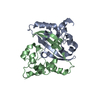 6yunS S: Starting model for refinement C: citing same article ( |
|---|---|
| Similar structure data |
-Links
 PDBj
PDBj


- Assembly
| Deposited unit | 
| ||||||||
|---|---|---|---|---|---|---|---|---|---|
| 1 | 
| ||||||||
| 2 | 
| ||||||||
| Unit cell |
|
-Components
| #1: Protein | Mass: 19569.055 Da / Num. of mol.: 4 / Fragment: C-terminal domain Source method: isolated from a genetically manipulated source Source: (gene. exp.) Severe acute respiratory syndrome coronavirus 2Production host:  #2: Chemical | ChemComp-SO4 / |   Mass: 96.063 Da / Num. of mol.: 1 / Source method: obtained synthetically / Formula: SO4 Mass: 96.063 Da / Num. of mol.: 1 / Source method: obtained synthetically / Formula: SO4#3: Chemical | 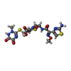  Mass: 554.580 Da / Num. of mol.: 3 / Source method: isolated from a natural source / Formula: C18H18N8O7S3 / Feature type: SUBJECT OF INVESTIGATION / Comment: antibiotic*YM Mass: 554.580 Da / Num. of mol.: 3 / Source method: isolated from a natural source / Formula: C18H18N8O7S3 / Feature type: SUBJECT OF INVESTIGATION / Comment: antibiotic*YM#4: Chemical | ChemComp-PEG / |   Mass: 106.120 Da / Num. of mol.: 1 / Source method: obtained synthetically / Formula: C4H10O3 Mass: 106.120 Da / Num. of mol.: 1 / Source method: obtained synthetically / Formula: C4H10O3#5: Water | ChemComp-HOH / |  Mass: 18.015 Da / Num. of mol.: 669 / Source method: isolated from a natural source / Formula: H2O Mass: 18.015 Da / Num. of mol.: 669 / Source method: isolated from a natural source / Formula: H2OHas ligand of interest | Y | Has protein modification | N | |
|---|
-Experimental details
-Experiment
| Experiment | Method: X-RAY DIFFRACTION / Number of used crystals: 1 |
|---|
- Sample preparation
| Crystal | Density Matthews: 2.67 Å3/Da / Density % sol: 53.85 % |
|---|---|
| Crystal grow | Temperature: 293 K / Method: vapor diffusion, sitting drop Details: 30% PEG4000, 0.2 M Lithium sulfate, 50 mM Tris pH 8.0 |
-Data collection
| Diffraction | Mean temperature: 100 K / Serial crystal experiment: N |
|---|---|
| Diffraction source | Source: ROTATING ANODE / Type: RIGAKU MICROMAX-007 HF / Wavelength: 1.54184 Å |
| Detector | Type: RIGAKU HyPix-6000HE / Detector: PIXEL / Date: Aug 24, 2022 |
| Radiation | Protocol: SINGLE WAVELENGTH / Monochromatic (M) / Laue (L): M / Scattering type: x-ray |
| Radiation wavelength | Wavelength: 1.54184 Å / Relative weight: 1 |
| Reflection | Resolution: 2→26.58 Å / Num. obs: 34801 / % possible obs: 99.2 % / Redundancy: 4.2 % / CC1/2: 0.997 / Net I/σ(I): 17.8 |
| Reflection shell | Resolution: 2→2.05 Å / Mean I/σ(I) obs: 3.8 / Num. unique obs: 2572 / CC1/2: 0.889 |
- Processing
| Software |
| ||||||||||||||||||||||||||||||||||||||||||||||||||||||||||||||||||||||||||||||||||||||||||||||||||||||||||||||||||||||||||||||||||||||||||||||||||||||||||||||||||||||||||||||||||||||
|---|---|---|---|---|---|---|---|---|---|---|---|---|---|---|---|---|---|---|---|---|---|---|---|---|---|---|---|---|---|---|---|---|---|---|---|---|---|---|---|---|---|---|---|---|---|---|---|---|---|---|---|---|---|---|---|---|---|---|---|---|---|---|---|---|---|---|---|---|---|---|---|---|---|---|---|---|---|---|---|---|---|---|---|---|---|---|---|---|---|---|---|---|---|---|---|---|---|---|---|---|---|---|---|---|---|---|---|---|---|---|---|---|---|---|---|---|---|---|---|---|---|---|---|---|---|---|---|---|---|---|---|---|---|---|---|---|---|---|---|---|---|---|---|---|---|---|---|---|---|---|---|---|---|---|---|---|---|---|---|---|---|---|---|---|---|---|---|---|---|---|---|---|---|---|---|---|---|---|---|---|---|---|---|
| Refinement | Method to determine structure: MOLECULAR REPLACEMENT Starting model: 6YUN Resolution: 2→26.58 Å / Cor.coef. Fo:Fc: 0.96 / Cor.coef. Fo:Fc free: 0.921 / SU B: 8.392 / SU ML: 0.104 / Cross valid method: THROUGHOUT / ESU R: 0.161 / ESU R Free: 0.153 / Stereochemistry target values: MAXIMUM LIKELIHOOD / Details: HYDROGENS HAVE BEEN ADDED IN THE RIDING POSITIONS
| ||||||||||||||||||||||||||||||||||||||||||||||||||||||||||||||||||||||||||||||||||||||||||||||||||||||||||||||||||||||||||||||||||||||||||||||||||||||||||||||||||||||||||||||||||||||
| Solvent computation | Ion probe radii: 0.8 Å / Shrinkage radii: 0.8 Å / VDW probe radii: 1.2 Å / Solvent model: MASK | ||||||||||||||||||||||||||||||||||||||||||||||||||||||||||||||||||||||||||||||||||||||||||||||||||||||||||||||||||||||||||||||||||||||||||||||||||||||||||||||||||||||||||||||||||||||
| Displacement parameters | Biso mean: 16.983 Å2
| ||||||||||||||||||||||||||||||||||||||||||||||||||||||||||||||||||||||||||||||||||||||||||||||||||||||||||||||||||||||||||||||||||||||||||||||||||||||||||||||||||||||||||||||||||||||
| Refinement step | Cycle: 1 / Resolution: 2→26.58 Å
| ||||||||||||||||||||||||||||||||||||||||||||||||||||||||||||||||||||||||||||||||||||||||||||||||||||||||||||||||||||||||||||||||||||||||||||||||||||||||||||||||||||||||||||||||||||||
| Refine LS restraints |
|