 Movie
Movie Controller

[English] 日本語
 Yorodumi
- PDB-8zci: X-ray structure of the human heart fatty acid-binding protein com... -
+ Open data

- Basic information
| Entry | Database: PDB / ID: 8zci | |||||||||||||||
|---|---|---|---|---|---|---|---|---|---|---|---|---|---|---|---|---|
| Title | X-ray structure of the human heart fatty acid-binding protein complexed with R-Ibuprofen | |||||||||||||||
 Components Components | Fatty acid-binding protein, heart | |||||||||||||||
 Keywords Keywords | LIPID BINDING PROTEIN / FABP / Complex / Binding protein / R-Ibuprofen | |||||||||||||||
| Function / homology |  Function and homology information Function and homology informationicosatetraenoic acid binding / positive regulation of long-chain fatty acid import into cell / regulation of phosphatidylcholine biosynthetic process / regulation of fatty acid oxidation / oleic acid binding / positive regulation of phospholipid biosynthetic process / intracellular lipid transport / response to fatty acid / phospholipid homeostasis / long-chain fatty acid transmembrane transporter activity ...icosatetraenoic acid binding / positive regulation of long-chain fatty acid import into cell / regulation of phosphatidylcholine biosynthetic process / regulation of fatty acid oxidation / oleic acid binding / positive regulation of phospholipid biosynthetic process / intracellular lipid transport / response to fatty acid / phospholipid homeostasis / long-chain fatty acid transmembrane transporter activity / long-chain fatty acid binding / Triglyceride catabolism / sarcoplasm / long-chain fatty acid transport / cytoskeletal protein binding / brown fat cell differentiation / cholesterol homeostasis / fatty acid metabolic process / response to insulin / response to xenobiotic stimulus / negative regulation of cell population proliferation / : / extracellular exosome / nucleus / cytosol Similarity search - Function | |||||||||||||||
| Biological species |  Homo sapiens (human) Homo sapiens (human) | |||||||||||||||
| Method |  X-RAY DIFFRACTION / SYNCHROTRON / MOLECULAR REPLACEMENT / Resolution: 1.35 Å X-RAY DIFFRACTION / SYNCHROTRON / MOLECULAR REPLACEMENT / Resolution: 1.35 Å | |||||||||||||||
 Authors Authors | Sugiyama, S. / Tanaka, J. / Matsuoka, S. / Murata, M. | |||||||||||||||
| Funding support |  Japan, 4items Japan, 4items
| |||||||||||||||
 Citation Citation | Journal: To Be Published Title: X-ray structure of the human heart fatty acid-binding protein complexed with R-Ibuprofen Authors: Sugiyama, S. / Tanaka, J. / Matsuoka, S. / Murata, M. | |||||||||||||||
| History |
|
- Structure visualization
| Structure viewer | Molecule: MolmilJmol/JSmol |
|---|
- Downloads & links
-Download
| PDBx/mmCIF format | 8zci.cif.gz | 85 KB | Display | PDBx/mmCIF format |
|---|---|---|---|---|
| PDB format | pdb8zci.ent.gz | 62.1 KB | Display | PDB format |
| PDBx/mmJSON format | 8zci.json.gz | Tree view | PDBx/mmJSON format | |
| Others |  Other downloads Other downloads |
-Validation report
| Arichive directory | https://data.pdbj.org/pub/pdb/validation_reports/zc/8zciftp://data.pdbj.org/pub/pdb/validation_reports/zc/8zci | HTTPS FTP |
|---|
-Related structure data
| Related structure data | 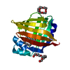 3wvmS S: Starting model for refinement |
|---|---|
| Similar structure data |
-Links
 PDBj
PDBj


- Assembly
| Deposited unit | 
| ||||||||
|---|---|---|---|---|---|---|---|---|---|
| 1 |
| ||||||||
| Unit cell |
|
-Components
-Protein , 1 types, 1 molecules A
| #1: Protein | Mass: 14879.022 Da / Num. of mol.: 1 Source method: isolated from a genetically manipulated source Source: (gene. exp.) Homo sapiens (human) / Gene: FABP3, FABP11, MDGI / Production host:  |
|---|
-Non-polymers , 7 types, 200 molecules 


| #2: Chemical | ChemComp-IZP / ( Mass: 206.281 Da / Num. of mol.: 1 / Source method: obtained synthetically / Formula: C13H18O2 / Feature type: SUBJECT OF INVESTIGATION Mass: 206.281 Da / Num. of mol.: 1 / Source method: obtained synthetically / Formula: C13H18O2 / Feature type: SUBJECT OF INVESTIGATION |
|---|---|
| #3: Chemical | ChemComp-PG4 /  Mass: 194.226 Da / Num. of mol.: 1 / Source method: obtained synthetically / Formula: C8H18O5 / Comment: precipitant*YM Mass: 194.226 Da / Num. of mol.: 1 / Source method: obtained synthetically / Formula: C8H18O5 / Comment: precipitant*YM |
| #4: Chemical | ChemComp-PEG /  Mass: 106.120 Da / Num. of mol.: 1 / Source method: obtained synthetically / Formula: C4H10O3 Mass: 106.120 Da / Num. of mol.: 1 / Source method: obtained synthetically / Formula: C4H10O3 |
| #5: Chemical | ChemComp-ACY /  Mass: 60.052 Da / Num. of mol.: 1 / Source method: obtained synthetically / Formula: C2H4O2 Mass: 60.052 Da / Num. of mol.: 1 / Source method: obtained synthetically / Formula: C2H4O2 |
| #6: Chemical | ChemComp-PGE /  Mass: 150.173 Da / Num. of mol.: 1 / Source method: obtained synthetically / Formula: C6H14O4 Mass: 150.173 Da / Num. of mol.: 1 / Source method: obtained synthetically / Formula: C6H14O4 |
| #7: Chemical | ChemComp-GOL /  Mass: 92.094 Da / Num. of mol.: 1 / Source method: obtained synthetically / Formula: C3H8O3 Mass: 92.094 Da / Num. of mol.: 1 / Source method: obtained synthetically / Formula: C3H8O3 |
| #8: Water | ChemComp-HOH / Mass: 18.015 Da / Num. of mol.: 194 / Source method: isolated from a natural source / Formula: H2O |
-Details
| Has ligand of interest | Y |
|---|---|
| Has protein modification | N |
-Experimental details
-Experiment
| Experiment | Method: X-RAY DIFFRACTION / Number of used crystals: 1 |
|---|
- Sample preparation
| Crystal | Density Matthews: 3.02 Å3/Da / Density % sol: 59.26 % |
|---|---|
| Crystal grow | Temperature: 293 K / Method: vapor diffusion, sitting drop / pH: 4.5 / Details: 0.1M Na-citrate (pH4.5), 40%PEG400 |
-Data collection
| Diffraction | Mean temperature: 100 K / Serial crystal experiment: N |
|---|---|
| Diffraction source | Source: SYNCHROTRON / Site: SPring-8 / Beamline: BL44XU / Wavelength: 0.9 Å |
| Detector | Type: MAR CCD 300 mm / Detector: CCD / Date: May 12, 2013 |
| Radiation | Protocol: SINGLE WAVELENGTH / Monochromatic (M) / Laue (L): M / Scattering type: x-ray |
| Radiation wavelength | Wavelength: 0.9 Å / Relative weight: 1 |
| Reflection | Resolution: 1.35→58.24 Å / Num. obs: 39678 / % possible obs: 99.3 % / Redundancy: 6.9 % / Rmerge(I) obs: 0.052 / Net I/σ(I): 12.4 |
| Reflection shell | Resolution: 1.35→1.37 Å / Rmerge(I) obs: 0.389 / Mean I/σ(I) obs: 2.5 / Num. unique obs: 1968 |
- Processing
| Software |
| ||||||||||||||||||||||||||||||||||||||||||||||||||||||||||||||||||||||||||||||||||||||||||||||||||||||||||||||||||||||||||||||||||||||||||||||||||||||||||||||||||||||||||||||||||||||
|---|---|---|---|---|---|---|---|---|---|---|---|---|---|---|---|---|---|---|---|---|---|---|---|---|---|---|---|---|---|---|---|---|---|---|---|---|---|---|---|---|---|---|---|---|---|---|---|---|---|---|---|---|---|---|---|---|---|---|---|---|---|---|---|---|---|---|---|---|---|---|---|---|---|---|---|---|---|---|---|---|---|---|---|---|---|---|---|---|---|---|---|---|---|---|---|---|---|---|---|---|---|---|---|---|---|---|---|---|---|---|---|---|---|---|---|---|---|---|---|---|---|---|---|---|---|---|---|---|---|---|---|---|---|---|---|---|---|---|---|---|---|---|---|---|---|---|---|---|---|---|---|---|---|---|---|---|---|---|---|---|---|---|---|---|---|---|---|---|---|---|---|---|---|---|---|---|---|---|---|---|---|---|---|
| Refinement | Method to determine structure: MOLECULAR REPLACEMENT Starting model: 3WVM Resolution: 1.35→31.93 Å / Cor.coef. Fo:Fc: 0.977 / Cor.coef. Fo:Fc free: 0.971 / SU B: 1.923 / SU ML: 0.033 / Cross valid method: THROUGHOUT / ESU R: 0.043 / ESU R Free: 0.044 / Stereochemistry target values: MAXIMUM LIKELIHOOD / Details: HYDROGENS HAVE BEEN ADDED IN THE RIDING POSITIONS
| ||||||||||||||||||||||||||||||||||||||||||||||||||||||||||||||||||||||||||||||||||||||||||||||||||||||||||||||||||||||||||||||||||||||||||||||||||||||||||||||||||||||||||||||||||||||
| Solvent computation | Ion probe radii: 0.8 Å / Shrinkage radii: 0.8 Å / VDW probe radii: 1.2 Å / Solvent model: MASK | ||||||||||||||||||||||||||||||||||||||||||||||||||||||||||||||||||||||||||||||||||||||||||||||||||||||||||||||||||||||||||||||||||||||||||||||||||||||||||||||||||||||||||||||||||||||
| Displacement parameters | Biso mean: 23.079 Å2
| ||||||||||||||||||||||||||||||||||||||||||||||||||||||||||||||||||||||||||||||||||||||||||||||||||||||||||||||||||||||||||||||||||||||||||||||||||||||||||||||||||||||||||||||||||||||
| Refinement step | Cycle: 1 / Resolution: 1.35→31.93 Å
| ||||||||||||||||||||||||||||||||||||||||||||||||||||||||||||||||||||||||||||||||||||||||||||||||||||||||||||||||||||||||||||||||||||||||||||||||||||||||||||||||||||||||||||||||||||||
| Refine LS restraints |
|