 Movie
Movie Controller

[English] 日本語
 Yorodumi
- PDB-8z96: Crystal structure of CrtAgo/TIR-APAZ in complex with guide DNA an... -
+ Open data

- Basic information
| Entry | Database: PDB / ID: 8z96 | ||||||
|---|---|---|---|---|---|---|---|
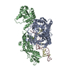
| Title | Crystal structure of CrtAgo/TIR-APAZ in complex with guide DNA and 21-nt target DNA | ||||||
 Components Components |
| ||||||
 Keywords Keywords | DNA BINDING PROTEIN/DNA / NADase / DNA / Complex / DNA BINDING PROTEIN-DNA complex | ||||||
| Function / homology |  Function and homology information Function and homology information | ||||||
| Biological species |  Thermoflavifilum thermophilum (bacteria) Thermoflavifilum thermophilum (bacteria) | ||||||
| Method |  X-RAY DIFFRACTION / SYNCHROTRON / MOLECULAR REPLACEMENT / Resolution: 3.36 Å X-RAY DIFFRACTION / SYNCHROTRON / MOLECULAR REPLACEMENT / Resolution: 3.36 Å | ||||||
 Authors Authors | Hu, R. / Chen, J. / Liu, L. | ||||||
| Funding support |  China, 1items China, 1items
| ||||||
 Citation Citation | Journal: Nucleic Acids Res. / Year: 2025 Title: Structural basis of ssDNA-guided NADase activation of prokaryotic SPARTA system. Authors: Hu, R. / Guo, C. / Liu, X. / Lin, Y. / Yang, Z. / Li, Z. / Yang, Y. / Ma, E. / Li, Y. / Chen, J. / Liu, L. | ||||||
| History |
|
- Structure visualization
| Structure viewer | Molecule: MolmilJmol/JSmol |
|---|
- Downloads & links
-Download
| PDBx/mmCIF format | 8z96.cif.gz | 226.6 KB | Display | PDBx/mmCIF format |
|---|---|---|---|---|
| PDB format | pdb8z96.ent.gz | 171.2 KB | Display | PDB format |
| PDBx/mmJSON format | 8z96.json.gz | Tree view | PDBx/mmJSON format | |
| Others |  Other downloads Other downloads |
-Validation report
| Arichive directory | https://data.pdbj.org/pub/pdb/validation_reports/z9/8z96ftp://data.pdbj.org/pub/pdb/validation_reports/z9/8z96 | HTTPS FTP |
|---|
-Related structure data


-Links
 PDBj
PDBj


- Assembly
| Deposited unit | 
| ||||||||||||
|---|---|---|---|---|---|---|---|---|---|---|---|---|---|
| 1 |
| ||||||||||||
| Unit cell |
| ||||||||||||
| Components on special symmetry positions |
|
-Components
-Protein , 2 types, 2 molecules AB
| #1: Protein | Mass: 58304.848 Da / Num. of mol.: 1 Source method: isolated from a genetically manipulated source Source: (gene. exp.) Thermoflavifilum thermophilum (bacteria)Gene: SAMN05660895_1671 / Production host: |
|---|---|
| #2: Protein | Mass: 49906.344 Da / Num. of mol.: 1 Source method: isolated from a genetically manipulated source Source: (gene. exp.) Thermoflavifilum thermophilum (bacteria)Gene: SAMN05660895_1670 / Production host: |
-DNA chain , 2 types, 2 molecules CD
| #3: DNA chain | Mass: 6588.266 Da / Num. of mol.: 1 / Source method: obtained synthetically / Source: (synth.) Thermoflavifilum thermophilum (bacteria) |
|---|---|
| #4: DNA chain | Mass: 6286.101 Da / Num. of mol.: 1 / Source method: obtained synthetically / Source: (synth.) Thermoflavifilum thermophilum (bacteria) |
-Non-polymers , 2 types, 26 molecules 

| #5: Chemical | ChemComp-MG /  Mass: 24.305 Da / Num. of mol.: 1 / Source method: isolated from a natural source / Formula: Mg Mass: 24.305 Da / Num. of mol.: 1 / Source method: isolated from a natural source / Formula: Mg |
|---|---|
| #6: Water | ChemComp-HOH / Mass: 18.015 Da / Num. of mol.: 25 / Source method: isolated from a natural source / Formula: H2O |
-Details
| Has ligand of interest | N |
|---|---|
| Has protein modification | N |
-Experimental details
-Experiment
| Experiment | Method: X-RAY DIFFRACTION / Number of used crystals: 1 |
|---|
- Sample preparation
| Crystal | Density Matthews: 3.56 Å3/Da / Density % sol: 65.42 % |
|---|---|
| Crystal grow | Temperature: 289 K / Method: vapor diffusion, hanging drop Details: 2% v/v Tacsimate, pH 7.0, 5% v/v 2-Propanol, 0.1 M Imidazole, pH 7.0, 8% w/v Polyethylene glycol 3350 |
-Data collection
| Diffraction | Mean temperature: 100 K / Serial crystal experiment: N |
|---|---|
| Diffraction source | Source: SYNCHROTRON / Site: SSRF / Beamline: BL18U1 / Wavelength: 0.94138 Å |
| Detector | Type: DECTRIS PILATUS3 6M / Detector: PIXEL / Date: Jan 30, 2024 |
| Radiation | Protocol: SINGLE WAVELENGTH / Monochromatic (M) / Laue (L): M / Scattering type: x-ray |
| Radiation wavelength | Wavelength: 0.94138 Å / Relative weight: 1 |
| Reflection | Resolution: 3.36→50 Å / Num. obs: 25189 / % possible obs: 100 % / Redundancy: 17.3 % / Biso Wilson estimate: 51.11 Å2 / Rpim(I) all: 0.052 / Net I/σ(I): 14.8 |
| Reflection shell | Resolution: 3.36→3.42 Å / Redundancy: 14.8 % / Mean I/σ(I) obs: 3 / Num. unique obs: 1220 / Rpim(I) all: 0.276 / % possible all: 100 |
- Processing
| Software |
| ||||||||||||||||||||||||||||||||||||||||||||||||||||||||
|---|---|---|---|---|---|---|---|---|---|---|---|---|---|---|---|---|---|---|---|---|---|---|---|---|---|---|---|---|---|---|---|---|---|---|---|---|---|---|---|---|---|---|---|---|---|---|---|---|---|---|---|---|---|---|---|---|---|
| Refinement | Method to determine structure: MOLECULAR REPLACEMENT / Resolution: 3.36→20.68 Å / SU ML: 0.4364 / Cross valid method: FREE R-VALUE / σ(F): 1.44 / Phase error: 28.5046 Stereochemistry target values: GeoStd + Monomer Library + CDL v1.2
| ||||||||||||||||||||||||||||||||||||||||||||||||||||||||
| Solvent computation | Shrinkage radii: 0.9 Å / VDW probe radii: 1.11 Å / Solvent model: FLAT BULK SOLVENT MODEL | ||||||||||||||||||||||||||||||||||||||||||||||||||||||||
| Displacement parameters | Biso mean: 59.48 Å2 | ||||||||||||||||||||||||||||||||||||||||||||||||||||||||
| Refinement step | Cycle: LAST / Resolution: 3.36→20.68 Å
| ||||||||||||||||||||||||||||||||||||||||||||||||||||||||
| Refine LS restraints |
| ||||||||||||||||||||||||||||||||||||||||||||||||||||||||
| LS refinement shell |
|
