 Movie
Movie Controller

[English] 日本語
 Yorodumi
- PDB-8z4d: Structure of the S-ring region of the Vibrio flagellar MS-ring pr... -
+ Open data

- Basic information
| Entry | Database: PDB / ID: 8z4d | |||||||||||||||||||||||||||||||||||||||||||||
|---|---|---|---|---|---|---|---|---|---|---|---|---|---|---|---|---|---|---|---|---|---|---|---|---|---|---|---|---|---|---|---|---|---|---|---|---|---|---|---|---|---|---|---|---|---|---|
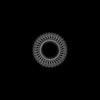
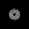
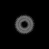
| Title | Structure of the S-ring region of the Vibrio flagellar MS-ring protein FliF with 34-fold symmetry applied | |||||||||||||||||||||||||||||||||||||||||||||
 Components Components | Flagellar M-ring protein,Flagellar motor switch protein FliG | |||||||||||||||||||||||||||||||||||||||||||||
 Keywords Keywords | MOTOR PROTEIN / Complex / Rotor | |||||||||||||||||||||||||||||||||||||||||||||
| Function / homology |  Function and homology information Function and homology informationbacterial-type flagellum basal body, MS ring / bacterial-type flagellum basal body / cytoskeletal motor activity / bacterial-type flagellum-dependent cell motility / chemotaxis / plasma membrane Similarity search - Function | |||||||||||||||||||||||||||||||||||||||||||||
| Biological species |  Vibrio alginolyticus (bacteria) Vibrio alginolyticus (bacteria) | |||||||||||||||||||||||||||||||||||||||||||||
| Method | ELECTRON MICROSCOPY / single particle reconstruction / cryo EM / Resolution: 3.33 Å | |||||||||||||||||||||||||||||||||||||||||||||
 Authors Authors | Takekawa, N. / Nishikino, T. / Kishikawa, J. / Hirose, M. / Kato, T. / Imada, K. / Homma, M. | |||||||||||||||||||||||||||||||||||||||||||||
| Funding support |  Japan, 4items Japan, 4items
| |||||||||||||||||||||||||||||||||||||||||||||
 Citation Citation | Journal: mBio / Year: 2024 Title: Structural analysis of S-ring composed of FliFG fusion proteins in marine polar flagellar motor. Authors: Norihiro Takekawa / Tatsuro Nishikino / Jun-Ichi Kishikawa / Mika Hirose / Miki Kinoshita / Seiji Kojima / Tohru Minamino / Takayuki Uchihashi / Takayuki Kato / Katsumi Imada / Michio Homma / Abstract: The marine bacterium possesses a polar flagellum driven by a sodium ion flow. The main components of the flagellar motor are the stator and rotor. The C-ring and MS-ring, which are composed of FliG ...The marine bacterium possesses a polar flagellum driven by a sodium ion flow. The main components of the flagellar motor are the stator and rotor. The C-ring and MS-ring, which are composed of FliG and FliF, respectively, are parts of the rotor. Here, we purified an MS-ring composed of FliF-FliG fusion proteins and solved the near-atomic resolution structure of the S-ring-the upper part of the MS-ring-using cryo-electron microscopy. This is the first report of an S-ring structure from , whereas, previously, only those from have been reported. The S-ring structure reveals novel features compared with that of , such as tilt angle differences of the RBM3 domain and the β-collar region, which contribute to the vertical arrangement of the upper part of the β-collar region despite the diversity in the RBM3 domain angles. Additionally, there is a decrease of the inter-subunit interaction between RBM3 domains, which influences the efficiency of the MS-ring formation in different bacterial species. Furthermore, although the inner-surface electrostatic properties of and S-rings are altered, the residues potentially interacting with other flagellar components, such as FliE and FlgB, are well structurally conserved in the S-ring. These comparisons clarified the conserved and non-conserved structural features of the MS-ring across different species.IMPORTANCEUnderstanding the structure and function of the flagellar motor in bacterial species is essential for uncovering the mechanisms underlying bacterial motility and pathogenesis. Our study revealed the structure of the S-ring, a part of its polar flagellar motor, and highlighted its unique features compared with the well-studied S-ring. The observed differences in the inter-subunit interactions and in the tilt angles between the and S-rings highlighted the species-specific variations and the mechanism for the optimization of MS-ring formation in the flagellar assembly. By concentrating on the region where the S-ring and the rod proteins interact, we uncovered conserved residues essential for the interaction. Our research contributes to the advancement of bacterial flagellar biology. | |||||||||||||||||||||||||||||||||||||||||||||
| History |
|
- Structure visualization
| Structure viewer | Molecule: MolmilJmol/JSmol |
|---|
- Downloads & links
-Download
| PDBx/mmCIF format | 8z4d.cif.gz | 1.3 MB | Display | PDBx/mmCIF format |
|---|---|---|---|---|
| PDB format | pdb8z4d.ent.gz | 878.5 KB | Display | PDB format |
| PDBx/mmJSON format | 8z4d.json.gz | Tree view | PDBx/mmJSON format | |
| Others |  Other downloads Other downloads |
-Validation report
| Arichive directory | https://data.pdbj.org/pub/pdb/validation_reports/z4/8z4dftp://data.pdbj.org/pub/pdb/validation_reports/z4/8z4d | HTTPS FTP |
|---|
-Related structure data
| Related structure data |  39761MC  8z4gC M: map data used to model this data C: citing same article ( |
|---|---|
| Similar structure data |
-Links
 PDBj
PDBj
- Assembly
| Deposited unit | 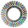
|
|---|---|
| 1 |
|
-Components
| #1: Protein | Mass: 104416.828 Da / Num. of mol.: 34 / Mutation: G214S Source method: isolated from a genetically manipulated source Source: (gene. exp.) Vibrio alginolyticus (bacteria) / Strain: 138-2 / Gene: fliF, Vag1382_20710, fliG, AL468_14360, JOS67_04865 / Production host: Has protein modification | N | |
|---|
-Experimental details
-Experiment
| Experiment | Method: ELECTRON MICROSCOPY |
|---|---|
| EM experiment | Aggregation state: PARTICLE / 3D reconstruction method: single particle reconstruction |
- Sample preparation
| Component | Name: Homomeric 34mer of FliFG fusion protein of Vibrio alginolyticus Type: COMPLEX / Entity ID: all / Source: RECOMBINANT | ||||||||||||||||||||
|---|---|---|---|---|---|---|---|---|---|---|---|---|---|---|---|---|---|---|---|---|---|
| Molecular weight | Value: 104 kDa/nm / Experimental value: NO | ||||||||||||||||||||
| Source (natural) | Organism: Vibrio alginolyticus (bacteria) / Strain: 138-2 | ||||||||||||||||||||
| Source (recombinant) | Organism: | ||||||||||||||||||||
| Buffer solution | pH: 8 | ||||||||||||||||||||
| Buffer component |
| ||||||||||||||||||||
| Specimen | Embedding applied: NO / Shadowing applied: NO / Staining applied: NO / Vitrification applied: YES | ||||||||||||||||||||
| Specimen support | Grid material: COPPER / Grid mesh size: 300 divisions/in. / Grid type: Quantifoil R1.2/1.3 | ||||||||||||||||||||
| Vitrification | Instrument: FEI VITROBOT MARK IV / Cryogen name: ETHANE / Humidity: 100 % / Chamber temperature: 277 K |
- Electron microscopy imaging
| Experimental equipment |  Model: Titan Krios / Image courtesy: FEI Company |
|---|---|
| Microscopy | Model: FEI TITAN KRIOS |
| Electron gun | Electron source:  FIELD EMISSION GUN / Accelerating voltage: 300 kV / Illumination mode: FLOOD BEAM FIELD EMISSION GUN / Accelerating voltage: 300 kV / Illumination mode: FLOOD BEAM |
| Electron lens | Mode: BRIGHT FIELD / Nominal magnification: 64000 X / Nominal defocus max: 1700 nm / Nominal defocus min: 700 nm / Alignment procedure: COMA FREE |
| Specimen holder | Cryogen: NITROGEN / Specimen holder model: FEI TITAN KRIOS AUTOGRID HOLDER |
| Image recording | Average exposure time: 7.3 sec. / Electron dose: 49.7 e/Å2 / Film or detector model: GATAN K3 BIOCONTINUUM (6k x 4k) / Num. of real images: 6372 |
| EM imaging optics | Energyfilter name: GIF Bioquantum / Energyfilter slit width: 20 eV |
- Processing
| EM software |
| |||||||||||||||||||||||||||
|---|---|---|---|---|---|---|---|---|---|---|---|---|---|---|---|---|---|---|---|---|---|---|---|---|---|---|---|---|
| CTF correction | Type: NONE | |||||||||||||||||||||||||||
| Particle selection | Num. of particles selected: 2059366 | |||||||||||||||||||||||||||
| Symmetry | Point symmetry: C34 (34 fold cyclic) | |||||||||||||||||||||||||||
| 3D reconstruction | Resolution: 3.33 Å / Resolution method: FSC 0.143 CUT-OFF / Num. of particles: 43546 / Symmetry type: POINT |
