Movie
Movie Controller

[English] 日本語
 Yorodumi
- EMDB-39765: Homomeric 35mer of the Vibrio flagellar MS-ring protein FliF with... -
+ Open data

- Basic information
| Entry |  | |||||||||||||||
|---|---|---|---|---|---|---|---|---|---|---|---|---|---|---|---|---|
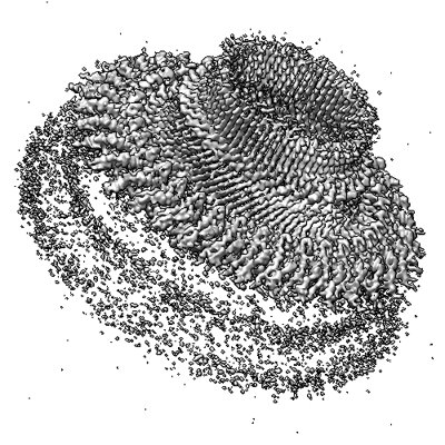
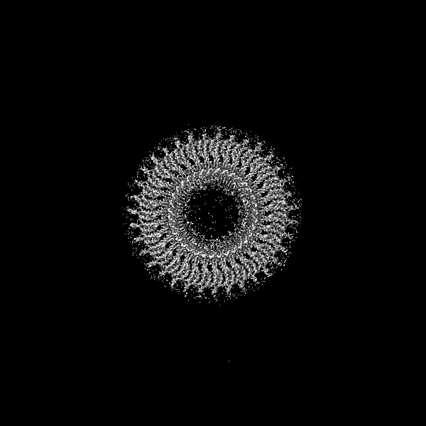
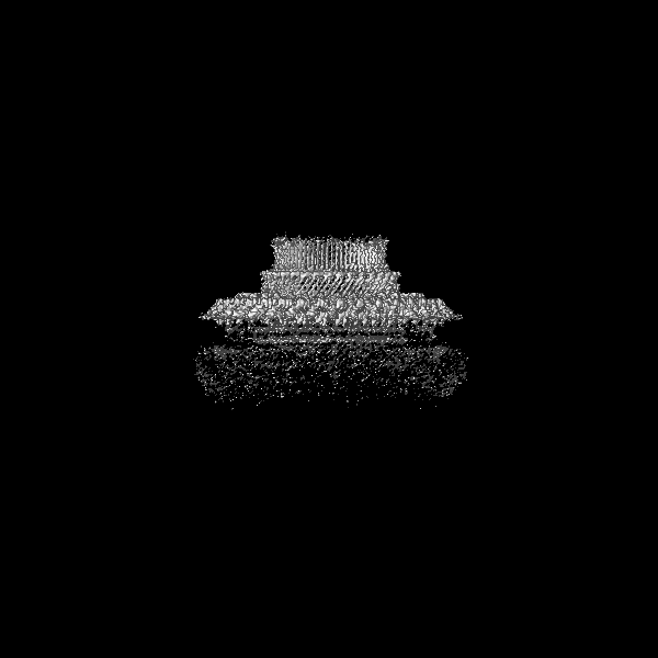
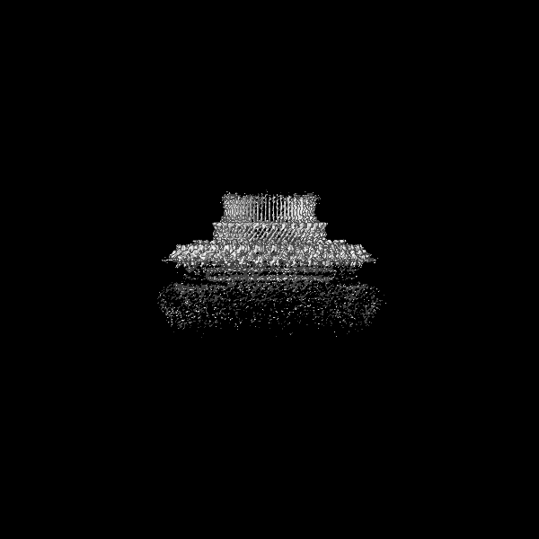
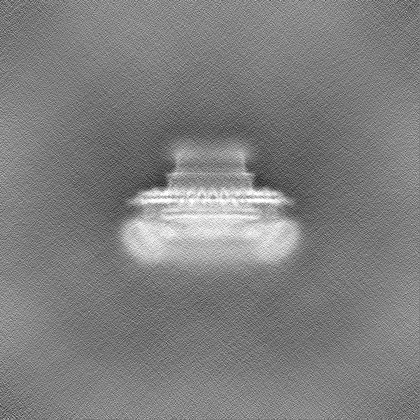
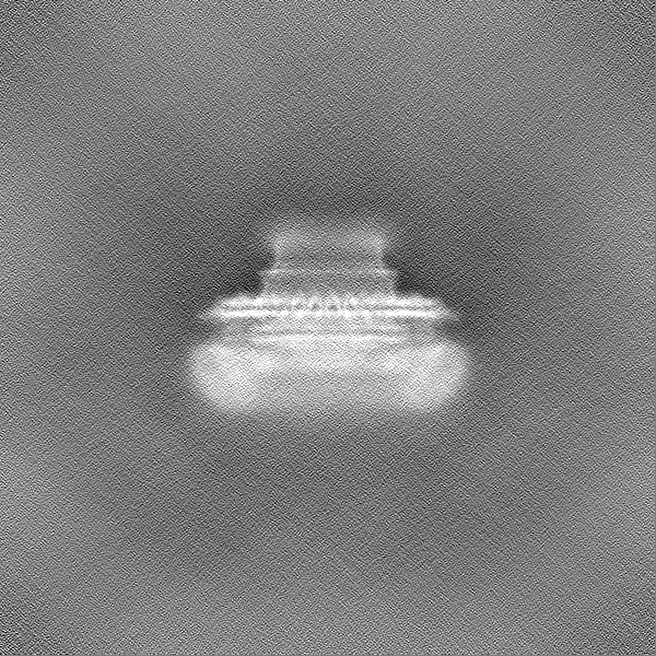
| Title | Homomeric 35mer of the Vibrio flagellar MS-ring protein FliF without symmetry imposition | |||||||||||||||
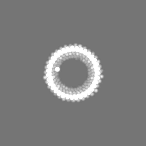
 Map data Map data | ||||||||||||||||
 Sample Sample |
| |||||||||||||||
 Keywords Keywords | Complex / Rotor / MOTOR PROTEIN | |||||||||||||||
| Biological species |  Vibrio alginolyticus (bacteria) Vibrio alginolyticus (bacteria) | |||||||||||||||
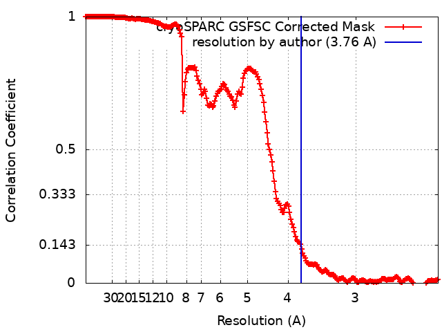
| Method | single particle reconstruction / cryo EM / Resolution: 3.76 Å | |||||||||||||||
 Authors Authors | Takekawa N / Nishikino T / Kishikawa J / Hirose M / Kato T / Imada K / Homma M | |||||||||||||||
| Funding support |  Japan, 4 items Japan, 4 items
| |||||||||||||||
 Citation Citation | Journal: mBio / Year: 2024 Title: Structural analysis of S-ring composed of FliFG fusion proteins in marine polar flagellar motor. Authors: Norihiro Takekawa / Tatsuro Nishikino / Jun-Ichi Kishikawa / Mika Hirose / Miki Kinoshita / Seiji Kojima / Tohru Minamino / Takayuki Uchihashi / Takayuki Kato / Katsumi Imada / Michio Homma / Abstract: The marine bacterium possesses a polar flagellum driven by a sodium ion flow. The main components of the flagellar motor are the stator and rotor. The C-ring and MS-ring, which are composed of FliG ...The marine bacterium possesses a polar flagellum driven by a sodium ion flow. The main components of the flagellar motor are the stator and rotor. The C-ring and MS-ring, which are composed of FliG and FliF, respectively, are parts of the rotor. Here, we purified an MS-ring composed of FliF-FliG fusion proteins and solved the near-atomic resolution structure of the S-ring-the upper part of the MS-ring-using cryo-electron microscopy. This is the first report of an S-ring structure from , whereas, previously, only those from have been reported. The S-ring structure reveals novel features compared with that of , such as tilt angle differences of the RBM3 domain and the β-collar region, which contribute to the vertical arrangement of the upper part of the β-collar region despite the diversity in the RBM3 domain angles. Additionally, there is a decrease of the inter-subunit interaction between RBM3 domains, which influences the efficiency of the MS-ring formation in different bacterial species. Furthermore, although the inner-surface electrostatic properties of and S-rings are altered, the residues potentially interacting with other flagellar components, such as FliE and FlgB, are well structurally conserved in the S-ring. These comparisons clarified the conserved and non-conserved structural features of the MS-ring across different species.IMPORTANCEUnderstanding the structure and function of the flagellar motor in bacterial species is essential for uncovering the mechanisms underlying bacterial motility and pathogenesis. Our study revealed the structure of the S-ring, a part of its polar flagellar motor, and highlighted its unique features compared with the well-studied S-ring. The observed differences in the inter-subunit interactions and in the tilt angles between the and S-rings highlighted the species-specific variations and the mechanism for the optimization of MS-ring formation in the flagellar assembly. By concentrating on the region where the S-ring and the rod proteins interact, we uncovered conserved residues essential for the interaction. Our research contributes to the advancement of bacterial flagellar biology. | |||||||||||||||
| History |
|
- Structure visualization
| Supplemental images |
|---|
- Downloads & links
-EMDB archive
| Map data | emd_39765.map.gz | 776.7 MB |  EMDB map data format EMDB map data format | |
|---|---|---|---|---|
| Header (meta data) | emd-39765-v30.xmlemd-39765.xml | 18.1 KB 18.1 KB | Display Display | EMDB header |
| FSC (resolution estimation) | emd_39765_fsc.xml | 19.8 KB | Display | FSC data file |
| Images |  emd_39765.png emd_39765.png | 200.7 KB | ||
| Masks | emd_39765_msk_1.map | 824 MB | Mask map | |
| Filedesc metadata | emd-39765.cif.gz | 5.6 KB | ||
| Others | emd_39765_half_map_1.map.gzemd_39765_half_map_2.map.gz | 763.2 MB 763.2 MB | ||
| Archive directory |  http://ftp.pdbj.org/pub/emdb/structures/EMD-39765ftp://ftp.pdbj.org/pub/emdb/structures/EMD-39765 http://ftp.pdbj.org/pub/emdb/structures/EMD-39765ftp://ftp.pdbj.org/pub/emdb/structures/EMD-39765 | HTTPS FTP |
-Related structure data


-Links
| EMDB pages | EMDB (EBI/PDBe) / EMDataResource |
|---|
-Map
| File | Download / File: emd_39765.map.gz / Format: CCP4 / Size: 824 MB / Type: IMAGE STORED AS FLOATING POINT NUMBER (4 BYTES) | ||||||||||||||||||||||||||||||||||||
|---|---|---|---|---|---|---|---|---|---|---|---|---|---|---|---|---|---|---|---|---|---|---|---|---|---|---|---|---|---|---|---|---|---|---|---|---|---|
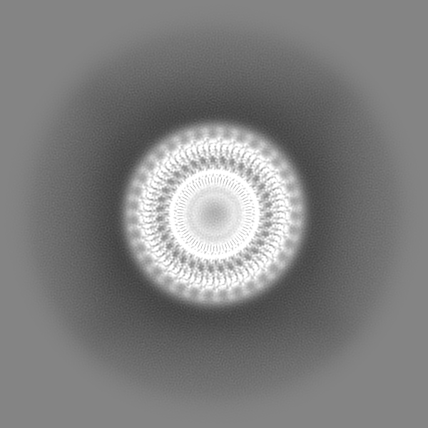
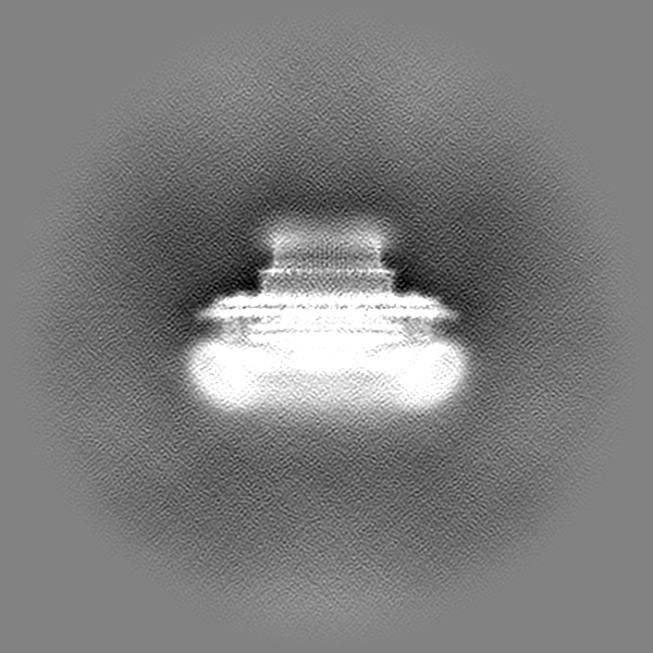
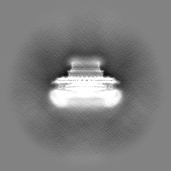
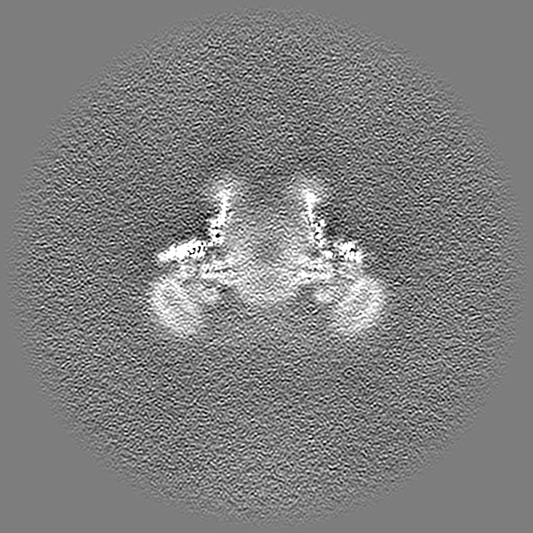
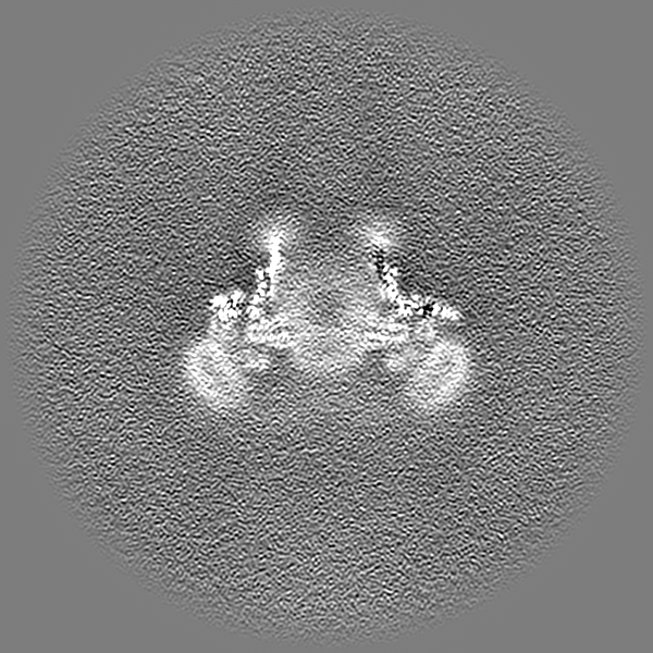
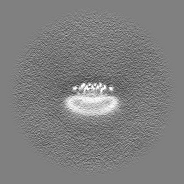
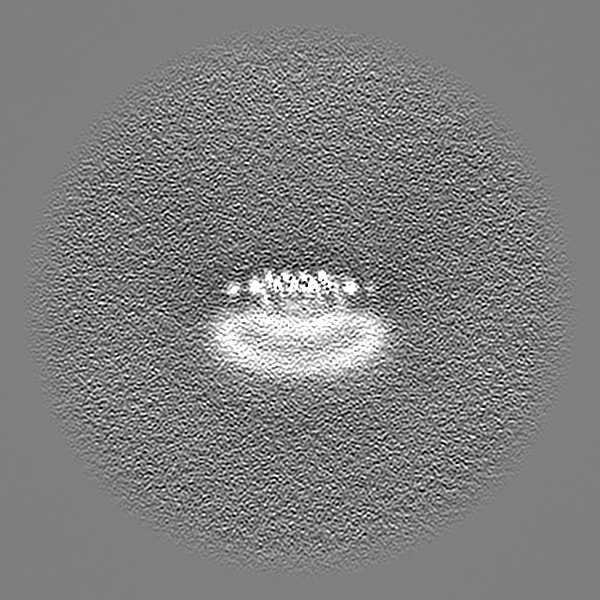
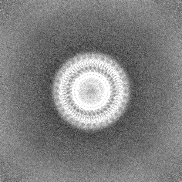
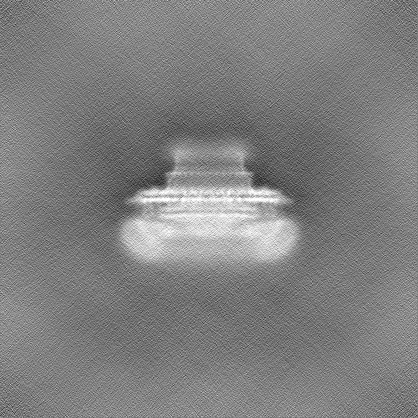
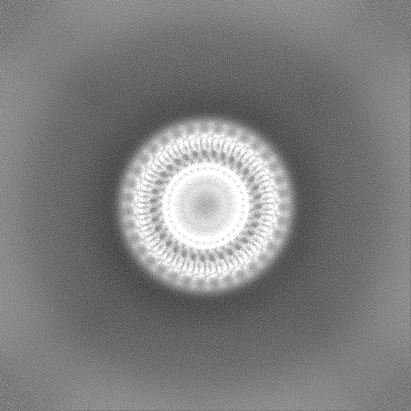
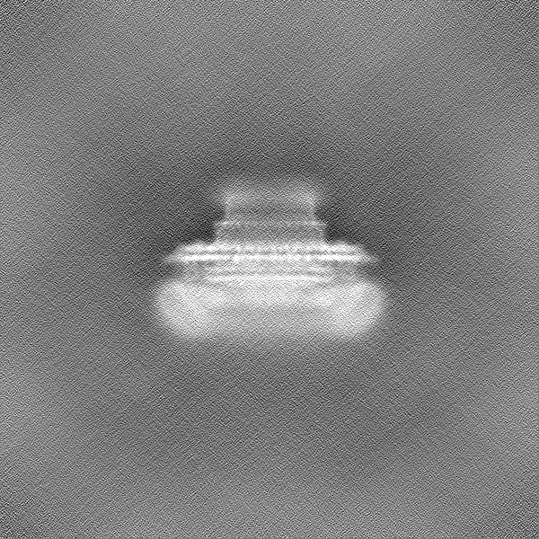
| Projections & slices | Image control
Images are generated by Spider. | ||||||||||||||||||||||||||||||||||||
| Voxel size | X=Y=Z: 1.14 Å | ||||||||||||||||||||||||||||||||||||
| Density |
| ||||||||||||||||||||||||||||||||||||
| Symmetry | Space group: 1 | ||||||||||||||||||||||||||||||||||||
| Details | EMDB XML:
|
 Z (Sec.)
 Y (Row.)
 X (Col.)


-Supplemental data
-Mask #1
| File | emd_39765_msk_1.map | ||||||||||||
|---|---|---|---|---|---|---|---|---|---|---|---|---|---|
| Projections & Slices |
| ||||||||||||
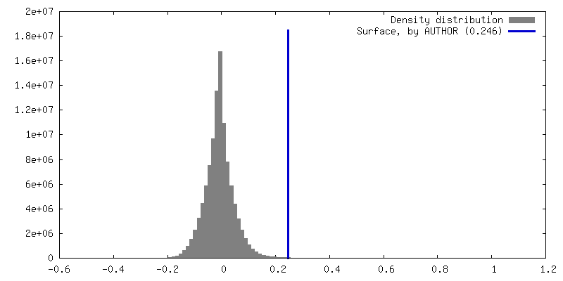
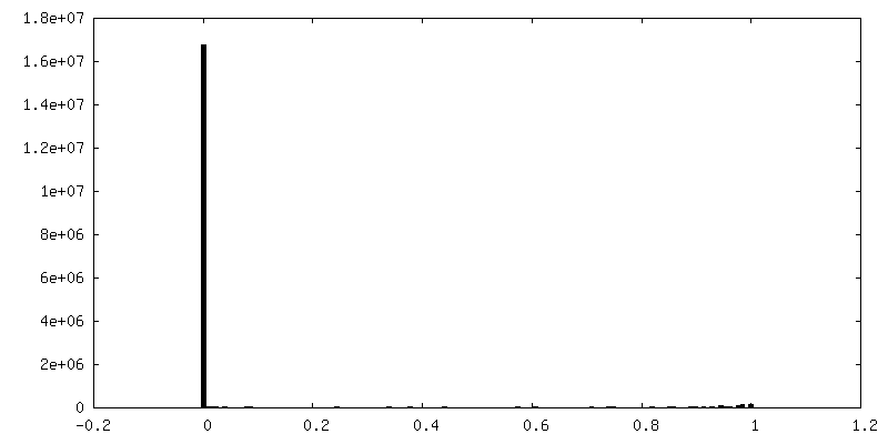
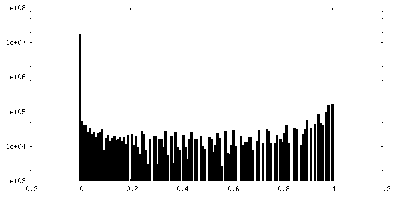
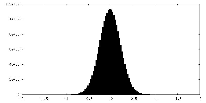
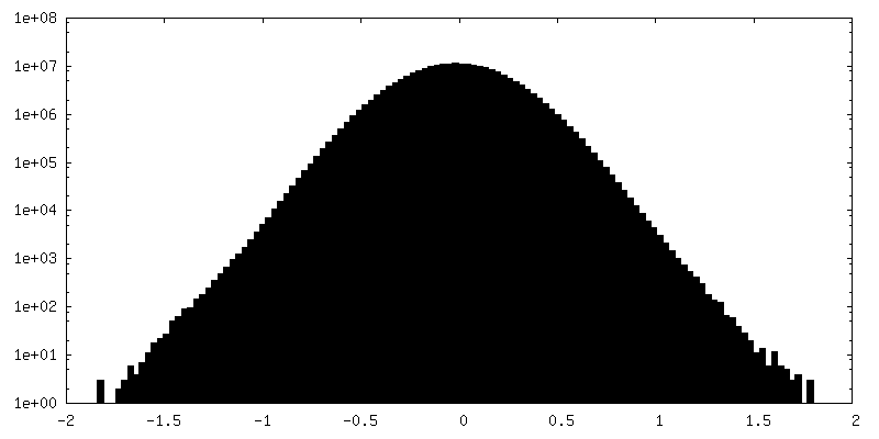
| Density Histograms |


-Half map: #1
| File | emd_39765_half_map_1.map | ||||||||||||
|---|---|---|---|---|---|---|---|---|---|---|---|---|---|
| Projections & Slices |
| ||||||||||||
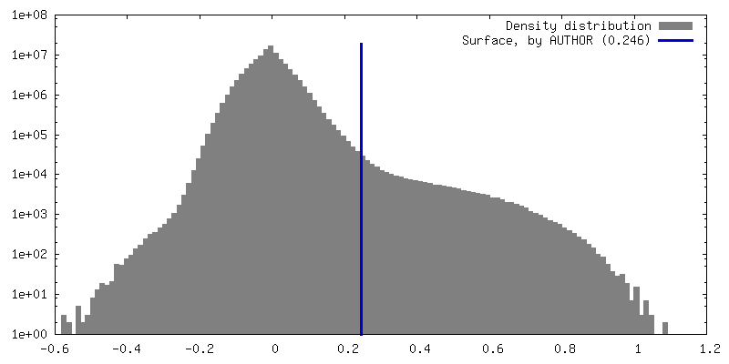
| Density Histograms |
-Half map: #2
| File | emd_39765_half_map_2.map | ||||||||||||
|---|---|---|---|---|---|---|---|---|---|---|---|---|---|
| Projections & Slices |
| ||||||||||||
| Density Histograms |

- Sample components
-Entire : Homomeric 35mer of FliFG fusion protein of Vibrio alginolyticus
| Entire | Name: Homomeric 35mer of FliFG fusion protein of Vibrio alginolyticus |
|---|---|
| Components |
|
-Supramolecule #1: Homomeric 35mer of FliFG fusion protein of Vibrio alginolyticus
| Supramolecule | Name: Homomeric 35mer of FliFG fusion protein of Vibrio alginolyticus type: complex / ID: 1 / Parent: 0 / Macromolecule list: all |
|---|---|
| Source (natural) | Organism: Vibrio alginolyticus (bacteria) / Strain: 138-2 |
| Molecular weight | Theoretical: 104 kDa/nm |
-Macromolecule #1: FliFG fusion protein
| Macromolecule | Name: FliFG fusion protein / type: protein_or_peptide / ID: 1 / Enantiomer: LEVO |
|---|---|
| Sequence | String: MNHKVHHHHH HIEGRHMADK STDLTVTEGG SDGALVASSD VDVESQNPDL EERSASKFDM AVGDLDLLRQ VVLVLSISIC VALIVMLFFW VKEPEMRPLG AYETEELIPV LDYLDQQKIN YKLDGNTISV ESSEYNSIKL GMVRSGVNQA TEAGDDILLQ DMGFGVSQRL ...String: MNHKVHHHHH HIEGRHMADK STDLTVTEGG SDGALVASSD VDVESQNPDL EERSASKFDM AVGDLDLLRQ VVLVLSISIC VALIVMLFFW VKEPEMRPLG AYETEELIPV LDYLDQQKIN YKLDGNTISV ESSEYNSIKL GMVRSGVNQA TEAGDDILLQ DMGFGVSQRL EQERLKLSRE RQLAQAIEEM KQVRKARVLL ALPKHSVFVR HNQEASASVF LTLSTGTNLK QQEVDSIVDM VASAVPGMKT SRITVTDQHG RLLSSGSQDP ASAARRKEQE LERSQEQALR EKIDSVLLPI LGYGNYTAQV DIQMDFSAVE QTRKRFDPNT PATRSEYALE DYNNGNMVAG IPGALSNQPP ADASIPQDVA QMKDGSVMGQ GSVRKESTRN FELDTTISHE RKQTGTVARQ TVSVAIKDRR QVNPDTGEVT YTPMSESEIN AIRQVLIGTV GFDQGRGDLL NVLSVKFAEP EAEQLEEPPI WEHPNFSDWV RWFASALVII VVVLVLVRPA MKKLLNPTSD DEDEMYGPDG LPIGADGETS LIGSDIESSE LFEFGSSIDL PNLHKDEDVL KAVRALVANE PELAAQVVKN WMNDMANDIV PQDDNAAGMP VELDVSTITG EEKAAILLLS LNEQDAAGII RHLEPKQVQR VGSAMAKAKD LSQDKVSAVH RAFLEDIQKY TNIGMGSEDF MRNALVAALG EDKANNLVDQ ILLGTGSKGL DSLKWMDPRQ VASIIVNEHP QIQTIVLSYL EADQSAEILS QFPERVRLDL MMRIANLEEV QPSALAELNE IMEKQFAGQA GAQAAKISGL KAAAEIMNYL DNNVEGLLME QIRDQDEDMA TQIQDLMFVF ENLVEVDDQG IQKLLRDVPQ DVLQKALKGA DDSLREKVFK NMSKRAAEMM RDDIEAMPPV RVADVEAAQK EILAIARRMA DAGELMLSGG ADEFL |
-Experimental details
-Structure determination
| Method | cryo EM |
|---|---|
 Processing Processing | single particle reconstruction |
| Aggregation state | particle |
-Sample preparation
| Buffer | pH: 8 Component:
| ||||||||||||
|---|---|---|---|---|---|---|---|---|---|---|---|---|---|
| Grid | Model: Quantifoil R1.2/1.3 / Material: COPPER / Mesh: 300 / Pretreatment - Type: GLOW DISCHARGE / Pretreatment - Atmosphere: AIR | ||||||||||||
| Vitrification | Cryogen name: ETHANE / Chamber humidity: 100 % / Chamber temperature: 277 K / Instrument: FEI VITROBOT MARK IV |
- Electron microscopy
| Microscope | FEI TITAN KRIOS |
|---|---|
| Specialist optics | Energy filter - Name: GIF Bioquantum / Energy filter - Slit width: 20 eV |
| Image recording | Film or detector model: GATAN K3 BIOCONTINUUM (6k x 4k) / Number real images: 6372 / Average exposure time: 7.3 sec. / Average electron dose: 49.7 e/Å2 |
| Electron beam | Acceleration voltage: 300 kV / Electron source:  FIELD EMISSION GUN FIELD EMISSION GUN |
| Electron optics | Illumination mode: FLOOD BEAM / Imaging mode: BRIGHT FIELD / Nominal defocus max: 1.7 µm / Nominal defocus min: 0.7000000000000001 µm / Nominal magnification: 64000 |
| Sample stage | Specimen holder model: FEI TITAN KRIOS AUTOGRID HOLDER / Cooling holder cryogen: NITROGEN |
| Experimental equipment |  Model: Titan Krios / Image courtesy: FEI Company |