Movie
Movie Controller


+ Open data

- Basic information
| Entry | Database: PDB / ID: 8yyv | ||||||
|---|---|---|---|---|---|---|---|
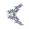
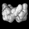
| Title | A dimeric STAT1-DNA complex | ||||||
 Components Components |
| ||||||
 Keywords Keywords | IMMUNE SYSTEM / innate immunity | ||||||
| Function / homology |  Function and homology information Function and homology informationmetanephric mesenchymal cell differentiation / negative regulation of metanephric nephron tubule epithelial cell differentiation / negative regulation by virus of viral protein levels in host cell / renal tubule development / ISGF3 complex / response to interferon-beta / metanephric mesenchymal cell proliferation involved in metanephros development / interleukin-27-mediated signaling pathway / negative regulation of mesenchymal to epithelial transition involved in metanephros morphogenesis / CCR5 chemokine receptor binding ...metanephric mesenchymal cell differentiation / negative regulation of metanephric nephron tubule epithelial cell differentiation / negative regulation by virus of viral protein levels in host cell / renal tubule development / ISGF3 complex / response to interferon-beta / metanephric mesenchymal cell proliferation involved in metanephros development / interleukin-27-mediated signaling pathway / negative regulation of mesenchymal to epithelial transition involved in metanephros morphogenesis / CCR5 chemokine receptor binding / Interleukin-9 signaling / Interleukin-21 signaling / interleukin-7-mediated signaling pathway / interleukin-9-mediated signaling pathway / Signaling by cytosolic FGFR1 fusion mutants / tumor necrosis factor receptor binding / Interleukin-27 signaling / Interleukin-35 Signalling / cell surface receptor signaling pathway via STAT / blood circulation / positive regulation of mesenchymal cell proliferation / NOTCH3 Intracellular Domain Regulates Transcription / Interleukin-20 family signaling / Interleukin-6 signaling / type I interferon-mediated signaling pathway / histone acetyltransferase binding / negative regulation of endothelial cell proliferation / ubiquitin-like protein ligase binding / positive regulation of interferon-alpha production / Regulation of IFNA/IFNB signaling / response to type II interferon / response to cAMP / cell surface receptor signaling pathway via JAK-STAT / response to mechanical stimulus / type II interferon-mediated signaling pathway / Regulation of IFNG signaling / Growth hormone receptor signaling / RNA polymerase II core promoter sequence-specific DNA binding / Signaling by PDGFRA transmembrane, juxtamembrane and kinase domain mutants / Signaling by PDGFRA extracellular domain mutants / cellular response to interferon-beta / Signaling by CSF3 (G-CSF) / positive regulation of defense response to virus by host / response to cytokine / response to nutrient / negative regulation of canonical NF-kappaB signal transduction / positive regulation of smooth muscle cell proliferation / Downstream signal transduction / Signaling by phosphorylated juxtamembrane, extracellular and kinase domain KIT mutants / negative regulation of angiogenesis / positive regulation of erythrocyte differentiation / protein phosphatase 2A binding / Downregulation of SMAD2/3:SMAD4 transcriptional activity / Turbulent (oscillatory, disturbed) flow shear stress activates signaling by PIEZO1 and integrins in endothelial cells / tumor necrosis factor-mediated signaling pathway / transcription corepressor binding / RNA polymerase II transcription regulatory region sequence-specific DNA binding / Inactivation of CSF3 (G-CSF) signaling / promoter-specific chromatin binding / ISG15 antiviral mechanism / response to hydrogen peroxide / Signaling by SCF-KIT / defense response / response to peptide hormone / PKR-mediated signaling / Signaling by CSF1 (M-CSF) in myeloid cells / cellular response to type II interferon / RNA polymerase II transcription regulator complex / Regulation of RUNX2 expression and activity / Interferon alpha/beta signaling / transcription coactivator binding / cellular response to insulin stimulus / Signaling by ALK fusions and activated point mutants / Interferon gamma signaling / positive regulation of nitric oxide biosynthetic process / regulation of cell population proliferation / double-stranded DNA binding / Interleukin-4 and Interleukin-13 signaling / histone binding / regulation of apoptotic process / defense response to virus / DNA-binding transcription factor activity, RNA polymerase II-specific / cadherin binding / RNA polymerase II cis-regulatory region sequence-specific DNA binding / response to xenobiotic stimulus / DNA-binding transcription factor activity / axon / dendrite / regulation of transcription by RNA polymerase II / chromatin / positive regulation of DNA-templated transcription / nucleolus / SARS-CoV-2 activates/modulates innate and adaptive immune responses / perinuclear region of cytoplasm / enzyme binding / negative regulation of transcription by RNA polymerase II / protein homodimerization activity / positive regulation of transcription by RNA polymerase II / protein-containing complex / nucleoplasm Similarity search - Function | ||||||
| Biological species |  Homo sapiens (human) Homo sapiens (human) | ||||||
| Method | ELECTRON MICROSCOPY / single particle reconstruction / cryo EM / Resolution: 3.07 Å | ||||||
 Authors Authors | Sugiyama, A. / Minami, M. / Sugita, Y. / Ose, T. | ||||||
| Funding support |  Japan, 1items Japan, 1items
| ||||||
 Citation Citation | Journal: Sci Signal / Year: 2025 Title: Structural analysis reveals how tetrameric tyrosine-phosphorylated STAT1 is targeted by the rabies virus P-protein. Authors: Aoi Sugiyama / Miku Minami / Kaito Ugajin / Satomi Inaba-Inoue / Nana Yabuno / Yuichiro Takekawa / Sun Xiaomei / Shiho Takei / Mina Sasaki / Tomo Nomai / Xinxin Jiang / Shunsuke Kita / ...Authors: Aoi Sugiyama / Miku Minami / Kaito Ugajin / Satomi Inaba-Inoue / Nana Yabuno / Yuichiro Takekawa / Sun Xiaomei / Shiho Takei / Mina Sasaki / Tomo Nomai / Xinxin Jiang / Shunsuke Kita / Katsumi Maenaka / Mika Hirose / Min Yao / Paul R Gooley / Gregory W Moseley / Yukihiko Sugita / Toyoyuki Ose /  Abstract: Signal transducer and activator of transcription (STAT) family members mediate signaling in the Janus kinase (JAK)-STAT pathway and are activated by phosphorylation at a conserved tyrosine residue, ...Signal transducer and activator of transcription (STAT) family members mediate signaling in the Janus kinase (JAK)-STAT pathway and are activated by phosphorylation at a conserved tyrosine residue, resulting in dimerization through reciprocal interactions between the phosphotyrosine and a Src homology 2 (SH2) domain. Tyrosine-phosphorylated STAT (pY-STAT) then translocates to the nucleus to induce the expression of genes encoding antiviral proteins. Although the active and functional forms of STATs are conventionally considered to be dimers, STATs can undergo higher-order oligomerization, which is implicated in regulating transcriptional activity. We present the cryo-electron microscopy (cryo-EM) structure of the tetrameric form of intact pY-STAT1 in complex with DNA, which indicates that interactions between the amino-terminal domains (NTDs) of STAT1 induce oligomerization. The tetrameric structure revealed a compact conformation with a previously uncharacterized binding interface: Two DNA-bound dimers are twofold symmetrically aligned to transform into a tandem DNA-binding model without NTD dimer separation. Moreover, biochemical analyses indicated that the rabies virus P-protein selectively targeted tetrameric pY-STAT1. Combined with data showing which regions contribute to the interaction between pY-STAT1 and the P-protein, we constructed a binding model explaining how P recognizes the pY-STAT1 tetramer. These data provide insight into how pathogenic viruses target signaling pathways that mediate the host immune response. | ||||||
| History |
|
- Structure visualization
| Structure viewer | Molecule: MolmilJmol/JSmol |
|---|
- Downloads & links
-Download
| PDBx/mmCIF format | 8yyv.cif.gz | 158.9 KB | Display | PDBx/mmCIF format |
|---|---|---|---|---|
| PDB format | pdb8yyv.ent.gz | 113.4 KB | Display | PDB format |
| PDBx/mmJSON format | 8yyv.json.gz | Tree view | PDBx/mmJSON format | |
| Others |  Other downloads Other downloads |
-Validation report
| Summary document | 8yyv_validation.pdf.gz | 1.2 MB | Display | wwPDB validaton report |
|---|---|---|---|---|
| Full document | 8yyv_full_validation.pdf.gz | 1.2 MB | Display | |
| Data in XML | 8yyv_validation.xml.gz | 34.1 KB | Display | |
| Data in CIF | 8yyv_validation.cif.gz | 48.9 KB | Display | |
| Arichive directory | https://data.pdbj.org/pub/pdb/validation_reports/yy/8yyvftp://data.pdbj.org/pub/pdb/validation_reports/yy/8yyv | HTTPS FTP |
-Related structure data
| Related structure data | 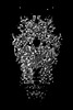 39680MC 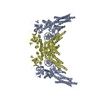 8yyuC M: map data used to model this data C: citing same article ( |
|---|---|
| Similar structure data |
-Links
 PDBj
PDBj


- Assembly
| Deposited unit | 
| ||||||||||||
|---|---|---|---|---|---|---|---|---|---|---|---|---|---|
| 1 | 
| ||||||||||||
| 2 |
| ||||||||||||
| 3 | 
| ||||||||||||
| Symmetry | Point symmetry: (Schoenflies symbol: C2 (2 fold cyclic)) | ||||||||||||
| Noncrystallographic symmetry (NCS) | NCS oper:
|
-Components
| #1: Protein | Mass: 90766.148 Da / Num. of mol.: 1 Source method: isolated from a genetically manipulated source Details: signal transducer and activator of transcription / Source: (gene. exp.) Homo sapiens (human) / Gene: STAT1 / Production host:  |
|---|---|
| #2: DNA chain | Mass: 5475.567 Da / Num. of mol.: 1 / Source method: obtained synthetically / Source: (synth.) Homo sapiens (human) |
| #3: DNA chain | Mass: 5555.615 Da / Num. of mol.: 1 / Source method: obtained synthetically / Source: (synth.) Homo sapiens (human) |
| Has ligand of interest | Y |
| Has protein modification | Y |
-Experimental details
-Experiment
| Experiment | Method: ELECTRON MICROSCOPY |
|---|---|
| EM experiment | Aggregation state: PARTICLE / 3D reconstruction method: single particle reconstruction |
- Sample preparation
| Component |
| ||||||||||||||||||||||||
|---|---|---|---|---|---|---|---|---|---|---|---|---|---|---|---|---|---|---|---|---|---|---|---|---|---|
| Molecular weight | Experimental value: NO | ||||||||||||||||||||||||
| Source (natural) | Organism: Homo sapiens (human) | ||||||||||||||||||||||||
| Source (recombinant) | Organism: | ||||||||||||||||||||||||
| Buffer solution | pH: 7.4 | ||||||||||||||||||||||||
| Specimen | Embedding applied: NO / Shadowing applied: NO / Staining applied: NO / Vitrification applied: YES | ||||||||||||||||||||||||
| Specimen support | Grid material: GOLD / Grid mesh size: 300 divisions/in. / Grid type: Quantifoil R0.6/1 | ||||||||||||||||||||||||
| Vitrification | Instrument: FEI VITROBOT MARK IV / Cryogen name: ETHANE / Humidity: 100 % / Chamber temperature: 4 K |
- Electron microscopy imaging
| Experimental equipment |  Model: Titan Krios / Image courtesy: FEI Company |
|---|---|
| Microscopy | Model: FEI TITAN KRIOS |
| Electron gun | Electron source:  FIELD EMISSION GUN / Accelerating voltage: 300 kV / Illumination mode: FLOOD BEAM FIELD EMISSION GUN / Accelerating voltage: 300 kV / Illumination mode: FLOOD BEAM |
| Electron lens | Mode: BRIGHT FIELD / Nominal magnification: 81000 X / Nominal defocus max: 18000 nm / Nominal defocus min: 800 nm / Cs: 0.032 mm / C2 aperture diameter: 50 µm |
| Specimen holder | Cryogen: NITROGEN / Specimen holder model: FEI TITAN KRIOS AUTOGRID HOLDER |
| Image recording | Electron dose: 50 e/Å2 / Film or detector model: GATAN K3 BIOQUANTUM (6k x 4k) |
| EM imaging optics | Energyfilter name: GIF Bioquantum / Energyfilter slit width: 20 eV Spherical aberration corrector: A Cs corrector (CEOS GmbH, Germany) |
- Processing
| EM software |
| ||||||||||||||||||||||||||||||||||||||||||||||||||||||||||||||||||||||||||||||||||||||||||||||||||||||||||
|---|---|---|---|---|---|---|---|---|---|---|---|---|---|---|---|---|---|---|---|---|---|---|---|---|---|---|---|---|---|---|---|---|---|---|---|---|---|---|---|---|---|---|---|---|---|---|---|---|---|---|---|---|---|---|---|---|---|---|---|---|---|---|---|---|---|---|---|---|---|---|---|---|---|---|---|---|---|---|---|---|---|---|---|---|---|---|---|---|---|---|---|---|---|---|---|---|---|---|---|---|---|---|---|---|---|---|---|
| CTF correction | Type: PHASE FLIPPING AND AMPLITUDE CORRECTION | ||||||||||||||||||||||||||||||||||||||||||||||||||||||||||||||||||||||||||||||||||||||||||||||||||||||||||
| Particle selection | Num. of particles selected: 4005846 | ||||||||||||||||||||||||||||||||||||||||||||||||||||||||||||||||||||||||||||||||||||||||||||||||||||||||||
| Symmetry | Point symmetry: C2 (2 fold cyclic) | ||||||||||||||||||||||||||||||||||||||||||||||||||||||||||||||||||||||||||||||||||||||||||||||||||||||||||
| 3D reconstruction | Resolution: 3.07 Å / Resolution method: FSC 0.143 CUT-OFF / Num. of particles: 115824 / Algorithm: BACK PROJECTION / Symmetry type: POINT | ||||||||||||||||||||||||||||||||||||||||||||||||||||||||||||||||||||||||||||||||||||||||||||||||||||||||||
| Atomic model building | Protocol: AB INITIO MODEL / Space: RECIPROCAL | ||||||||||||||||||||||||||||||||||||||||||||||||||||||||||||||||||||||||||||||||||||||||||||||||||||||||||
| Atomic model building | PDB-ID: 1BF5 Accession code: 1BF5 / Source name: PDB / Type: experimental model | ||||||||||||||||||||||||||||||||||||||||||||||||||||||||||||||||||||||||||||||||||||||||||||||||||||||||||
| Refinement | Resolution: 3.07→3.07 Å / Cor.coef. Fo:Fc: 0.943 / SU B: 7.494 / SU ML: 0.121 / ESU R: 0.135 Stereochemistry target values: MAXIMUM LIKELIHOOD WITH PHASES Details: HYDROGENS HAVE BEEN USED IF PRESENT IN THE INPUT
| ||||||||||||||||||||||||||||||||||||||||||||||||||||||||||||||||||||||||||||||||||||||||||||||||||||||||||
| Solvent computation | Solvent model: PARAMETERS FOR MASK CACLULATION | ||||||||||||||||||||||||||||||||||||||||||||||||||||||||||||||||||||||||||||||||||||||||||||||||||||||||||
| Displacement parameters | Biso mean: 113.477 Å2 | ||||||||||||||||||||||||||||||||||||||||||||||||||||||||||||||||||||||||||||||||||||||||||||||||||||||||||
| Refinement step | Cycle: 1 / Total: 5249 | ||||||||||||||||||||||||||||||||||||||||||||||||||||||||||||||||||||||||||||||||||||||||||||||||||||||||||
| Refine LS restraints |
|