 Movie
Movie Controller

[English] 日本語
 Yorodumi
- PDB-8yxk: X-ray structure of Clostridioides difficile endolysin Ecd09610 gl... -
+ Open data

- Basic information
| Entry | Database: PDB / ID: 8yxk | ||||||
|---|---|---|---|---|---|---|---|
| Title | X-ray structure of Clostridioides difficile endolysin Ecd09610 glucosaminidase domain. | ||||||
 Components Components | Phage cell wall hydrolase | ||||||
 Keywords Keywords | HYDROLASE / Clostridioides difficile / endolysin | ||||||
| Function / homology | : / Mannosyl-glycoprotein endo-beta-N-acetylglucosamidase-like domain / Mannosyl-glycoprotein endo-beta-N-acetylglucosaminidase / NlpC/P60 family / NlpC/P60 domain profile. / Endopeptidase, NLPC/P60 domain / amidase activity / Papain-like cysteine peptidase superfamily / Phage cell wall hydrolase Function and homology information Function and homology information | ||||||
| Biological species |  Clostridioides difficile (bacteria) Clostridioides difficile (bacteria) | ||||||
| Method |  X-RAY DIFFRACTION / MOLECULAR REPLACEMENT / Resolution: 1.87 Å X-RAY DIFFRACTION / MOLECULAR REPLACEMENT / Resolution: 1.87 Å | ||||||
 Authors Authors | Kamitori, S. / Tamai, E. | ||||||
| Funding support |  Japan, 1items Japan, 1items
| ||||||
 Citation Citation | Journal: Biochem.Biophys.Res.Commun. / Year: 2024 Title: X-ray structure and mutagenesis analyses of Clostridioides difficile endolysin Ecd09610 glucosaminidase domain. Authors: Sekiya, H. / Nonaka, Y. / Kamitori, S. / Miyaji, T. / Tamai, E. | ||||||
| History |
|
- Structure visualization
| Structure viewer | Molecule: MolmilJmol/JSmol |
|---|
- Downloads & links
-Download
| PDBx/mmCIF format | 8yxk.cif.gz | 75.2 KB | Display | PDBx/mmCIF format |
|---|---|---|---|---|
| PDB format | pdb8yxk.ent.gz | 52.6 KB | Display | PDB format |
| PDBx/mmJSON format | 8yxk.json.gz | Tree view | PDBx/mmJSON format | |
| Others |  Other downloads Other downloads |
-Validation report
| Arichive directory | https://data.pdbj.org/pub/pdb/validation_reports/yx/8yxkftp://data.pdbj.org/pub/pdb/validation_reports/yx/8yxk | HTTPS FTP |
|---|
-Related structure data

-Links
 PDBj
PDBj- Assembly
| Deposited unit | 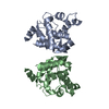
| ||||||||
|---|---|---|---|---|---|---|---|---|---|
| 1 | 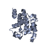
| ||||||||
| 2 | 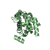
| ||||||||
| Unit cell |
|
-Components
| #1: Protein | Mass: 22397.062 Da / Num. of mol.: 2 / Fragment: glucosaminidase domain Source method: isolated from a genetically manipulated source Source: (gene. exp.) Clostridioides difficile (strain 630) (bacteria)Gene: CD630_09610, CD630_29030 / Production host: #2: Water | ChemComp-HOH / |  Mass: 18.015 Da / Num. of mol.: 80 / Source method: isolated from a natural source / Formula: H2O Mass: 18.015 Da / Num. of mol.: 80 / Source method: isolated from a natural source / Formula: H2O |
|---|
-Experimental details
-Experiment
| Experiment | Method: X-RAY DIFFRACTION / Number of used crystals: 1 |
|---|
- Sample preparation
| Crystal | Density Matthews: 2.06 Å3/Da / Density % sol: 40.22 % |
|---|---|
| Crystal grow | Temperature: 293 K / Method: vapor diffusion, sitting drop / pH: 9 / Details: 20% (w/v) PEG 3350, 200mM sodium thiocyanate |
-Data collection
| Diffraction | Mean temperature: 100 K / Serial crystal experiment: N |
|---|---|
| Diffraction source | Source: ROTATING ANODE / Type: RIGAKU MICROMAX-007 HF / Wavelength: 1.5418 Å |
| Detector | Type: RIGAKU RAXIS VII / Detector: IMAGE PLATE / Date: Jul 21, 2021 |
| Radiation | Protocol: SINGLE WAVELENGTH / Monochromatic (M) / Laue (L): M / Scattering type: x-ray |
| Radiation wavelength | Wavelength: 1.5418 Å / Relative weight: 1 |
| Reflection | Resolution: 1.87→19.322 Å / Num. obs: 29563 / % possible obs: 98.5 % / Redundancy: 3.7 % / CC1/2: 0.996 / Net I/σ(I): 9 |
| Reflection shell | Resolution: 1.87→1.92 Å / Num. unique obs: 2151 / CC1/2: 0.761 |
- Processing
| Software |
| ||||||||||||||||||||||||||||||||||||||||||||||||||||||||||||||||||||||||||||||||||||||||||||||||||||||||||||||||||||||||||||||||||||||||||||||||||||||
|---|---|---|---|---|---|---|---|---|---|---|---|---|---|---|---|---|---|---|---|---|---|---|---|---|---|---|---|---|---|---|---|---|---|---|---|---|---|---|---|---|---|---|---|---|---|---|---|---|---|---|---|---|---|---|---|---|---|---|---|---|---|---|---|---|---|---|---|---|---|---|---|---|---|---|---|---|---|---|---|---|---|---|---|---|---|---|---|---|---|---|---|---|---|---|---|---|---|---|---|---|---|---|---|---|---|---|---|---|---|---|---|---|---|---|---|---|---|---|---|---|---|---|---|---|---|---|---|---|---|---|---|---|---|---|---|---|---|---|---|---|---|---|---|---|---|---|---|---|---|---|---|
| Refinement | Method to determine structure: MOLECULAR REPLACEMENT / Resolution: 1.87→19.322 Å / Cor.coef. Fo:Fc: 0.912 / Cor.coef. Fo:Fc free: 0.886 / SU B: 4.573 / SU ML: 0.132 / Cross valid method: FREE R-VALUE / ESU R: 0.171 / ESU R Free: 0.161 Details: Hydrogens have been added in their riding positions
| ||||||||||||||||||||||||||||||||||||||||||||||||||||||||||||||||||||||||||||||||||||||||||||||||||||||||||||||||||||||||||||||||||||||||||||||||||||||
| Solvent computation | Ion probe radii: 0.8 Å / Shrinkage radii: 0.8 Å / VDW probe radii: 1.2 Å / Solvent model: MASK BULK SOLVENT | ||||||||||||||||||||||||||||||||||||||||||||||||||||||||||||||||||||||||||||||||||||||||||||||||||||||||||||||||||||||||||||||||||||||||||||||||||||||
| Displacement parameters | Biso mean: 21.628 Å2
| ||||||||||||||||||||||||||||||||||||||||||||||||||||||||||||||||||||||||||||||||||||||||||||||||||||||||||||||||||||||||||||||||||||||||||||||||||||||
| Refinement step | Cycle: LAST / Resolution: 1.87→19.322 Å
| ||||||||||||||||||||||||||||||||||||||||||||||||||||||||||||||||||||||||||||||||||||||||||||||||||||||||||||||||||||||||||||||||||||||||||||||||||||||
| Refine LS restraints |
| ||||||||||||||||||||||||||||||||||||||||||||||||||||||||||||||||||||||||||||||||||||||||||||||||||||||||||||||||||||||||||||||||||||||||||||||||||||||
| LS refinement shell |
|
