+ データを開く
- 基本情報
基本情報
| 登録情報 | データベース: PDB / ID: 8yhx | ||||||
|---|---|---|---|---|---|---|---|
| タイトル | Cryo-EM structure of the trimeric HerA | ||||||
 要素 要素 |
| ||||||
 キーワード キーワード | IMMUNE SYSTEM / antiphage system | ||||||
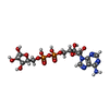
| 機能・相同性 | SIR2-like domain / ADENOSINE-5-DIPHOSPHORIBOSE / : / SIR2 family protein 機能・相同性情報 機能・相同性情報 | ||||||
| 生物種 |  | ||||||
| 手法 | 電子顕微鏡法 / 単粒子再構成法 / クライオ電子顕微鏡法 / 解像度: 2.81 Å | ||||||
 データ登録者 データ登録者 | Zhen, X. / Zhou, B. / Xiong, X. | ||||||
| 資金援助 |  中国, 1件 中国, 1件
| ||||||
 引用 引用 |  ジャーナル: Nat Commun / 年: 2024 ジャーナル: Nat Commun / 年: 2024タイトル: Mechanistic basis for the allosteric activation of NADase activity in the Sir2-HerA antiphage defense system. 著者: Xiangkai Zhen / Biao Zhou / Zihe Liu / Xurong Wang / Heyu Zhao / Shuxian Wu / Zekai Li / Jiamin Liang / Wanyue Zhang / Qingjian Zhu / Jun He / Xiaoli Xiong / Songying Ouyang /  要旨: Sir2-HerA is a widely distributed antiphage system composed of a RecA-like ATPase (HerA) and an effector with potential NADase activity (Sir2). Sir2-HerA is believed to provide defense against phage ...Sir2-HerA is a widely distributed antiphage system composed of a RecA-like ATPase (HerA) and an effector with potential NADase activity (Sir2). Sir2-HerA is believed to provide defense against phage infection in Sir2-dependent NAD depletion to arrest the growth of infected cells. However, the detailed mechanism underlying its antiphage activity remains largely unknown. Here, we report functional investigations of Sir2-HerA from Staphylococcus aureus (SaSir2-HerA), unveiling that the NADase function of SaSir2 can be allosterically activated by the binding of SaHerA, which then assembles into a supramolecular complex with NADase activity. By combining the cryo-EM structure of SaSir2-HerA in complex with the NAD cleavage product, it is surprisingly observed that Sir2 protomers that interact with HerA are in the activated state, which is due to the opening of the α15-helix covering the active site, allowing NAD to access the catalytic pocket for hydrolysis. In brief, our study provides a comprehensive view of an allosteric activation mechanism for Sir2 NADase activity in the Sir2-HerA immune system. | ||||||
| 履歴 |
|
- 構造の表示
構造の表示
| 構造ビューア | 分子:  Molmil Molmil Jmol/JSmol Jmol/JSmol |
|---|
- ダウンロードとリンク
- ダウンロード
| PDBx/mmCIF形式 |  8yhx.cif.gz 8yhx.cif.gz | 1.4 MB | 表示 |  PDBx/mmCIF形式 PDBx/mmCIF形式 |
|---|---|---|---|---|
| PDB形式 |  pdb8yhx.ent.gz pdb8yhx.ent.gz | 表示 |  PDB形式 PDB形式 | |
| PDBx/mmJSON形式 |  8yhx.json.gz 8yhx.json.gz | ツリー表示 |  PDBx/mmJSON形式 PDBx/mmJSON形式 | |
| その他 |  その他のダウンロード その他のダウンロード |
-検証レポート
| アーカイブディレクトリ |  https://data.pdbj.org/pub/pdb/validation_reports/yh/8yhx https://data.pdbj.org/pub/pdb/validation_reports/yh/8yhx ftp://data.pdbj.org/pub/pdb/validation_reports/yh/8yhx ftp://data.pdbj.org/pub/pdb/validation_reports/yh/8yhx | HTTPS FTP |
|---|
-関連構造データ
| 関連構造データ |  39302MC  8yhoC M: このデータのモデリングに利用したマップデータ C: 同じ文献を引用 ( |
|---|---|
| 類似構造データ | 類似検索 - 機能・相同性  F&H 検索 F&H 検索 |
- リンク
リンク
- 集合体
集合体
| 登録構造単位 | 
|
|---|---|
| 1 |
|
- 要素
要素
| #1: タンパク質 | 分子量: 65255.168 Da / 分子数: 6 / 由来タイプ: 組換発現 由来: (組換発現)  遺伝子: GO782_01940 / 発現宿主:  #2: タンパク質 | 分子量: 50710.613 Da / 分子数: 12 / 由来タイプ: 組換発現 由来: (組換発現)  遺伝子: GO782_01935 / 発現宿主:  #3: 化合物 | ChemComp-APR / 研究の焦点であるリガンドがあるか | Y | Has protein modification | N | |
|---|
-実験情報
-実験
| 実験 | 手法: 電子顕微鏡法 |
|---|---|
| EM実験 | 試料の集合状態: PARTICLE / 3次元再構成法: 単粒子再構成法 |
- 試料調製
試料調製
| 構成要素 | 名称: Cryo-Em structure of the trimeric HerA / タイプ: COMPLEX / Entity ID: #1-#2 / 由来: RECOMBINANT |
|---|---|
| 由来(天然) | 生物種:  |
| 由来(組換発現) | 生物種:  |
| 緩衝液 | pH: 7.5 |
| 試料 | 包埋: NO / シャドウイング: NO / 染色: NO / 凍結: YES |
| 急速凍結 | 凍結剤: ETHANE |
- 電子顕微鏡撮影
| 実験機器 |  モデル: Titan Krios / 画像提供: FEI Company |
|---|---|
| 顕微鏡 | モデル: TFS KRIOS |
| 電子銃 | 電子線源:  FIELD EMISSION GUN / 加速電圧: 300 kV / 照射モード: FLOOD BEAM FIELD EMISSION GUN / 加速電圧: 300 kV / 照射モード: FLOOD BEAM |
| 電子レンズ | モード: BRIGHT FIELD / 最大 デフォーカス(公称値): 2400 nm / 最小 デフォーカス(公称値): 800 nm |
| 撮影 | 電子線照射量: 50 e/Å2 フィルム・検出器のモデル: FEI FALCON IV (4k x 4k) |
- 解析
解析
| EMソフトウェア | 名称: PHENIX / バージョン: 1.13_2998: / カテゴリ: モデル精密化 |
|---|---|
| CTF補正 | タイプ: PHASE FLIPPING AND AMPLITUDE CORRECTION |
| 3次元再構成 | 解像度: 2.81 Å / 解像度の算出法: FSC 0.143 CUT-OFF / 粒子像の数: 79232 / 対称性のタイプ: POINT |
 ムービー
ムービー コントローラー


 PDBj
PDBj


