 Movie
Movie Controller


+ Open data

- Basic information
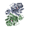
| Entry | Database: PDB / ID: 8yfp | ||||||
|---|---|---|---|---|---|---|---|
| Title | Chito oligosaccharide deacetylase from vibrio campbellii (VhCOD) | ||||||
 Components Components | NodB homology domain-containing protein | ||||||
 Keywords Keywords | HYDROLASE / Vibrio campbellii / Chito oligosaccharide deacetylase / Deacetylase Enzyme / CE4 family member | ||||||
| Function / homology |  Function and homology information Function and homology informationhydrolase activity, acting on carbon-nitrogen (but not peptide) bonds / hydrolase activity, hydrolyzing O-glycosyl compounds / carbohydrate binding / carbohydrate metabolic process / extracellular region / metal ion binding Similarity search - Function | ||||||
| Biological species |  Vibrio campbellii ATCC BAA-1116 (bacteria) Vibrio campbellii ATCC BAA-1116 (bacteria) | ||||||
| Method |  X-RAY DIFFRACTION / SYNCHROTRON / MOLECULAR REPLACEMENT / Resolution: 2.45 Å X-RAY DIFFRACTION / SYNCHROTRON / MOLECULAR REPLACEMENT / Resolution: 2.45 Å | ||||||
 Authors Authors | Sirikan, P. / Tamo, F. / Robinson, R.C. / Wipa, S. | ||||||
| Funding support |  Thailand, 1items Thailand, 1items
| ||||||
 Citation Citation | Journal: J.Biol.Chem. / Year: 2025 Title: Structure and loop dynamics of a chitooligosaccharide deacetylase from the marine bacterium Vibrio campbellii (harveyi). Authors: Pongnan, S. / Robinson, R.C. / Lampela, O. / Juffer, A. / Fukamizo, T. / Suginta, W. | ||||||
| History |
|
- Structure visualization
| Structure viewer | Molecule: MolmilJmol/JSmol |
|---|
- Downloads & links
-Download
| PDBx/mmCIF format | 8yfp.cif.gz | 327.8 KB | Display | PDBx/mmCIF format |
|---|---|---|---|---|
| PDB format | pdb8yfp.ent.gz | 267.9 KB | Display | PDB format |
| PDBx/mmJSON format | 8yfp.json.gz | Tree view | PDBx/mmJSON format | |
| Others |  Other downloads Other downloads |
-Validation report
| Arichive directory | https://data.pdbj.org/pub/pdb/validation_reports/yf/8yfpftp://data.pdbj.org/pub/pdb/validation_reports/yf/8yfp | HTTPS FTP |
|---|
-Related structure data


-Links
 PDBj
PDBj
- Assembly
| Deposited unit | 
| ||||||||
|---|---|---|---|---|---|---|---|---|---|
| 1 | 
| ||||||||
| 2 | 
| ||||||||
| Unit cell |
|
-Components
| #1: Protein | Mass: 45596.020 Da / Num. of mol.: 2 Source method: isolated from a genetically manipulated source Source: (gene. exp.) Vibrio campbellii ATCC BAA-1116 (bacteria)Gene: VIBHAR_03626 / Production host: #2: Chemical |   Mass: 65.409 Da / Num. of mol.: 2 / Source method: obtained synthetically / Formula: Zn / Feature type: SUBJECT OF INVESTIGATION Mass: 65.409 Da / Num. of mol.: 2 / Source method: obtained synthetically / Formula: Zn / Feature type: SUBJECT OF INVESTIGATION#3: Chemical |   Mass: 24.305 Da / Num. of mol.: 2 / Source method: obtained synthetically / Formula: Mg / Feature type: SUBJECT OF INVESTIGATION Mass: 24.305 Da / Num. of mol.: 2 / Source method: obtained synthetically / Formula: Mg / Feature type: SUBJECT OF INVESTIGATION#4: Chemical |   Mass: 40.078 Da / Num. of mol.: 3 / Source method: isolated from a natural source / Formula: Ca / Feature type: SUBJECT OF INVESTIGATION Mass: 40.078 Da / Num. of mol.: 3 / Source method: isolated from a natural source / Formula: Ca / Feature type: SUBJECT OF INVESTIGATION#5: Water | ChemComp-HOH / |  Mass: 18.015 Da / Num. of mol.: 243 / Source method: isolated from a natural source / Formula: H2O Mass: 18.015 Da / Num. of mol.: 243 / Source method: isolated from a natural source / Formula: H2OHas ligand of interest | Y | Has protein modification | Y | |
|---|
-Experimental details
-Experiment
| Experiment | Method: X-RAY DIFFRACTION / Number of used crystals: 1 |
|---|
- Sample preparation
| Crystal | Density Matthews: 4.28 Å3/Da / Density % sol: 70.58 % / Description: Hexagonal crytal |
|---|---|
| Crystal grow | Temperature: 292 K / Method: vapor diffusion, sitting drop / pH: 7.5 Details: 0.06M Divalent (0.3M Magnesium chloride hexahydrate, 0.3M Calcium chloride), 0.1M buffer system2 (1M Sodium HEPES, MOPS (acid) pH 7.5, 35% precipitant mix 4 (25% v/v MPD, 25% PEG 100, 25% w/v PEG 3350) |
-Data collection
| Diffraction | Mean temperature: 100 K / Ambient temp details: 100 / Serial crystal experiment: N |
|---|---|
| Diffraction source | Source: SYNCHROTRON / Site: NSRRC  / Beamline: TPS 05A / Wavelength: 0.9997 Å / Beamline: TPS 05A / Wavelength: 0.9997 Å |
| Detector | Type: DECTRIS EIGER2 S 9M / Detector: PIXEL / Date: Jun 18, 2021 |
| Radiation | Protocol: SINGLE WAVELENGTH / Monochromatic (M) / Laue (L): M / Scattering type: x-ray |
| Radiation wavelength | Wavelength: 0.9997 Å / Relative weight: 1 |
| Reflection | Resolution: 2.45→19.933 Å / Num. obs: 55208 / % possible obs: 99 % / Redundancy: 20 % / Biso Wilson estimate: 33.23 Å2 / CC1/2: 0.971 / CC star: 0.993 / Rmerge(I) obs: 0.203 / Rpim(I) all: 0.045 / Rrim(I) all: 0.208 / Net I/σ(I): 19 |
| Reflection shell | Resolution: 2.8→6.75 Å / Rmerge(I) obs: 2.041 / Num. unique obs: 37317 / CC1/2: 0.971 / CC star: 0.993 / Rpim(I) all: 0.182 / Rrim(I) all: 0.848 |
- Processing
| Software |
| |||||||||||||||||||||||||||||||||||||||||||||||||||||||||||||||||||||||||||||||||||||||||||||||||||||||||||||||||||||||||||||||||||||||||||||||||||
|---|---|---|---|---|---|---|---|---|---|---|---|---|---|---|---|---|---|---|---|---|---|---|---|---|---|---|---|---|---|---|---|---|---|---|---|---|---|---|---|---|---|---|---|---|---|---|---|---|---|---|---|---|---|---|---|---|---|---|---|---|---|---|---|---|---|---|---|---|---|---|---|---|---|---|---|---|---|---|---|---|---|---|---|---|---|---|---|---|---|---|---|---|---|---|---|---|---|---|---|---|---|---|---|---|---|---|---|---|---|---|---|---|---|---|---|---|---|---|---|---|---|---|---|---|---|---|---|---|---|---|---|---|---|---|---|---|---|---|---|---|---|---|---|---|---|---|---|---|
| Refinement | Method to determine structure: MOLECULAR REPLACEMENT / Resolution: 2.45→19.93 Å / SU ML: 0.25 / Cross valid method: FREE R-VALUE / σ(F): 1.34 / Phase error: 23.33 / Stereochemistry target values: ML
| |||||||||||||||||||||||||||||||||||||||||||||||||||||||||||||||||||||||||||||||||||||||||||||||||||||||||||||||||||||||||||||||||||||||||||||||||||
| Solvent computation | Shrinkage radii: 0.9 Å / VDW probe radii: 1.11 Å / Solvent model: FLAT BULK SOLVENT MODEL | |||||||||||||||||||||||||||||||||||||||||||||||||||||||||||||||||||||||||||||||||||||||||||||||||||||||||||||||||||||||||||||||||||||||||||||||||||
| Refinement step | Cycle: LAST / Resolution: 2.45→19.93 Å
| |||||||||||||||||||||||||||||||||||||||||||||||||||||||||||||||||||||||||||||||||||||||||||||||||||||||||||||||||||||||||||||||||||||||||||||||||||
| Refine LS restraints |
| |||||||||||||||||||||||||||||||||||||||||||||||||||||||||||||||||||||||||||||||||||||||||||||||||||||||||||||||||||||||||||||||||||||||||||||||||||
| LS refinement shell |
| |||||||||||||||||||||||||||||||||||||||||||||||||||||||||||||||||||||||||||||||||||||||||||||||||||||||||||||||||||||||||||||||||||||||||||||||||||
| Refinement TLS params. | Method: refined / Origin x: -10.1185 Å / Origin y: 28.892 Å / Origin z: -41.5108 Å
| |||||||||||||||||||||||||||||||||||||||||||||||||||||||||||||||||||||||||||||||||||||||||||||||||||||||||||||||||||||||||||||||||||||||||||||||||||
| Refinement TLS group | Selection details: all |
