 Movie
Movie Controller


+ Open data

- Basic information
| Entry | Database: PDB / ID: 8yfc | ||||||
|---|---|---|---|---|---|---|---|
| Title | Human PIEZO1-A1988V-MDFIC | ||||||
 Components Components |
| ||||||
 Keywords Keywords | MEMBRANE PROTEIN / Human PIEZO1 | ||||||
| Function / homology |  Function and homology information Function and homology informationmechanosensitive monoatomic cation channel activity / positive regulation of viral transcription / cuticular plate / positive regulation of integrin activation / detection of mechanical stimulus / positive regulation of cell-cell adhesion mediated by integrin / mechanosensitive monoatomic ion channel activity / stereocilium / Tat protein binding / regulation of Wnt signaling pathway ...mechanosensitive monoatomic cation channel activity / positive regulation of viral transcription / cuticular plate / positive regulation of integrin activation / detection of mechanical stimulus / positive regulation of cell-cell adhesion mediated by integrin / mechanosensitive monoatomic ion channel activity / stereocilium / Tat protein binding / regulation of Wnt signaling pathway / regulation of JNK cascade / negative regulation of protein import into nucleus / Mechanical load activates signaling by PIEZO1 and integrins in osteocytes / positive regulation of myotube differentiation / lamellipodium membrane / monoatomic cation transport / monoatomic cation channel activity / endoplasmic reticulum-Golgi intermediate compartment membrane / Turbulent (oscillatory, disturbed) flow shear stress activates signaling by PIEZO1 and integrins in endothelial cells / cyclin binding / cellular response to mechanical stimulus / regulation of membrane potential / High laminar flow shear stress activates signaling by PIEZO1 and PECAM1:CDH5:KDR in endothelial cells / DNA-binding transcription factor binding / negative regulation of DNA-templated transcription / endoplasmic reticulum membrane / positive regulation of DNA-templated transcription / nucleolus / endoplasmic reticulum / extracellular region / nucleus / plasma membrane / cytoplasm Similarity search - Function | ||||||
| Biological species |  Homo sapiens (human) Homo sapiens (human) | ||||||
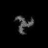
| Method | ELECTRON MICROSCOPY / single particle reconstruction / cryo EM / Resolution: 3.2 Å | ||||||
 Authors Authors | Zhang, M.F. | ||||||
| Funding support |  China, 1items China, 1items
| ||||||
 Citation Citation | Journal: Elife / Year: 2025 Title: Structure of human PIEZO1 and its slow-inactivating channelopathy mutants. Authors: Yuanyue Shan / Xinyi Guo / Mengmeng Zhang / Meiyu Chen / Ying Li / Mingfeng Zhang / Duanqing Pei / Abstract: PIEZO channels transmit mechanical force signals to cells, allowing them to make critical decisions during development and in pathophysiological conditions. Their fast/slow inactivation modes have ...PIEZO channels transmit mechanical force signals to cells, allowing them to make critical decisions during development and in pathophysiological conditions. Their fast/slow inactivation modes have been implicated in mechanopathologies but remain poorly understood. Here, we report several near-atomic resolution cryo-EM structures of fast-inactivating wild-type human PIEZO1 (hPIEZO1) and its slow-inactivating channelopathy mutants with or without its auxiliary subunit MDFIC. Our results suggest that hPIEZO1 has a more flattened and extended architecture than curved mouse PIEZO1 (mPIEZO1). The multi-lipidated MDFIC subunits insert laterally into the hPIEZO1 pore module like mPIEZO1, resulting in a more curved and extended state. Interestingly, the high-resolution structures suggest that the pore lipids, which directly seal the central hydrophobic pore, may be involved in the rapid inactivation of hPIEZO1. While the severe hereditary erythrocytosis mutant R2456H significantly slows down the inactivation of hPIEZO1, the hPIEZO1-R2456H-MDFIC complex shows a more curved and contracted structure with an inner helix twist due to the broken link between the pore lipid and R2456H. These results suggest that the pore lipids may be involved in the mechanopathological rapid inactivation mechanism of PIEZO channels. #1: Journal: Elife / Year: 2024Title: Structure of human PIEZO1 and its slow inactivating channelopathy mutants. Authors: Shan, Y. / Guo, X. / Zhang, M. / Chen, M. / Li, Y. / Zhang, M.F. / Pei, D. | ||||||
| History |
|
- Structure visualization
| Structure viewer | Molecule: MolmilJmol/JSmol |
|---|
- Downloads & links
-Download
| PDBx/mmCIF format | 8yfc.cif.gz | 781.4 KB | Display | PDBx/mmCIF format |
|---|---|---|---|---|
| PDB format | pdb8yfc.ent.gz | 598.5 KB | Display | PDB format |
| PDBx/mmJSON format | 8yfc.json.gz | Tree view | PDBx/mmJSON format | |
| Others |  Other downloads Other downloads |
-Validation report
| Arichive directory | https://data.pdbj.org/pub/pdb/validation_reports/yf/8yfcftp://data.pdbj.org/pub/pdb/validation_reports/yf/8yfc | HTTPS FTP |
|---|
-Related structure data
| Related structure data |  39219MC  8yezC  8yfgC 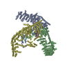 8zu3C  8zu8C M: map data used to model this data C: citing same article ( |
|---|---|
| Similar structure data |


-Links
 PDBj
PDBj
- Assembly
| Deposited unit | 
|
|---|---|
| 1 |
|
-Components
| #1: Protein | Mass: 287094.062 Da / Num. of mol.: 3 Source method: isolated from a genetically manipulated source Source: (gene. exp.) Homo sapiens (human) / Gene: PIEZO1, FAM38A, KIAA0233 / Production host: Homo sapiens (human) / References: UniProt: Q92508#2: Protein | Mass: 25814.113 Da / Num. of mol.: 3 Source method: isolated from a genetically manipulated source Source: (gene. exp.) Homo sapiens (human) / Gene: MDFIC / Production host: Homo sapiens (human) / References: UniProt: Q9P1T7#3: Chemical | ChemComp-D12 /   Mass: 170.335 Da / Num. of mol.: 15 / Source method: obtained synthetically / Formula: C12H26 / Feature type: SUBJECT OF INVESTIGATION Mass: 170.335 Da / Num. of mol.: 15 / Source method: obtained synthetically / Formula: C12H26 / Feature type: SUBJECT OF INVESTIGATION#4: Chemical |   Mass: 746.050 Da / Num. of mol.: 3 / Source method: obtained synthetically / Formula: C41H80NO8P / Feature type: SUBJECT OF INVESTIGATION Mass: 746.050 Da / Num. of mol.: 3 / Source method: obtained synthetically / Formula: C41H80NO8P / Feature type: SUBJECT OF INVESTIGATIONHas ligand of interest | Y | Has protein modification | Y | |
|---|
-Experimental details
-Experiment
| Experiment | Method: ELECTRON MICROSCOPY |
|---|---|
| EM experiment | Aggregation state: PARTICLE / 3D reconstruction method: single particle reconstruction |
- Sample preparation
| Component | Name: Human PIEZO1-A1988V-MDFIC / Type: COMPLEX / Entity ID: #1-#2 / Source: RECOMBINANT |
|---|---|
| Source (natural) | Organism: Homo sapiens (human) |
| Source (recombinant) | Organism: Homo sapiens (human) |
| Buffer solution | pH: 7.4 |
| Specimen | Embedding applied: NO / Shadowing applied: NO / Staining applied: NO / Vitrification applied: YES |
| Vitrification | Cryogen name: ETHANE |
- Electron microscopy imaging
| Microscopy | Model: FEI TECNAI 10 |
|---|---|
| Electron gun | Electron source:  FIELD EMISSION GUN / Accelerating voltage: 300 kV / Illumination mode: FLOOD BEAM FIELD EMISSION GUN / Accelerating voltage: 300 kV / Illumination mode: FLOOD BEAM |
| Electron lens | Mode: BRIGHT FIELD / Nominal defocus max: 2000 nm / Nominal defocus min: 1200 nm |
| Image recording | Electron dose: 40 e/Å2 / Film or detector model: FEI FALCON IV (4k x 4k) |
- Processing
| CTF correction | Type: NONE |
|---|---|
| 3D reconstruction | Resolution: 3.2 Å / Resolution method: FSC 0.143 CUT-OFF / Num. of particles: 40207 / Symmetry type: POINT |
