 Movie
Movie Controller

[English] 日本語
 Yorodumi
- PDB-8ybn: Crystal structure of nanobody SEB-Nb8 bound to staphylococcal ent... -
+ Open data

- Basic information
| Entry | Database: PDB / ID: 8ybn | ||||||||||||
|---|---|---|---|---|---|---|---|---|---|---|---|---|---|
| Title | Crystal structure of nanobody SEB-Nb8 bound to staphylococcal enterotoxin B (SEB) | ||||||||||||
 Components Components |
| ||||||||||||
 Keywords Keywords | ANTITOXIN / antibody / nanobody / toxin / staphylococcal enterotoxin B (SEB) | ||||||||||||
| Function / homology |  Function and homology information Function and homology information | ||||||||||||
| Biological species |   Staphylococcus aureus (bacteria) Staphylococcus aureus (bacteria) | ||||||||||||
| Method |  X-RAY DIFFRACTION / SYNCHROTRON / MOLECULAR REPLACEMENT / Resolution: 2.18 Å X-RAY DIFFRACTION / SYNCHROTRON / MOLECULAR REPLACEMENT / Resolution: 2.18 Å | ||||||||||||
 Authors Authors | Ding, Y. / Zong, X. / Liu, R. / Liu, P. | ||||||||||||
| Funding support |  China, 3items China, 3items
| ||||||||||||
 Citation Citation | Journal: Int.J.Biol.Macromol. / Year: 2024 Title: Structural insights into the binding of nanobodies to the Staphylococcal enterotoxin B. Authors: Zong, X. / Liu, P. / Wang, Z. / Zhu, H. / Zhong, C. / Zhong, P. / Jiang, H. / Liu, J. / Ma, Z. / Liu, X. / Liu, R. / Ding, Y. | ||||||||||||
| History |
|
- Structure visualization
| Structure viewer | Molecule: MolmilJmol/JSmol |
|---|
- Downloads & links
-Download
| PDBx/mmCIF format | 8ybn.cif.gz | 87.3 KB | Display | PDBx/mmCIF format |
|---|---|---|---|---|
| PDB format | pdb8ybn.ent.gz | 63.9 KB | Display | PDB format |
| PDBx/mmJSON format | 8ybn.json.gz | Tree view | PDBx/mmJSON format | |
| Others |  Other downloads Other downloads |
-Validation report
| Arichive directory | https://data.pdbj.org/pub/pdb/validation_reports/yb/8ybnftp://data.pdbj.org/pub/pdb/validation_reports/yb/8ybn | HTTPS FTP |
|---|
-Related structure data


-Links
 PDBj
PDBj


- Assembly
| Deposited unit | 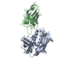
| ||||||||
|---|---|---|---|---|---|---|---|---|---|
| 1 |
| ||||||||
| Unit cell |
|
-Components
| #1: Protein | Mass: 28542.262 Da / Num. of mol.: 1 Source method: isolated from a genetically manipulated source Source: (gene. exp.) Staphylococcus aureus (bacteria) / Gene: entB / Production host: |
|---|---|
| #2: Antibody | Mass: 14427.987 Da / Num. of mol.: 1 Source method: isolated from a genetically manipulated source Source: (gene. exp.) |
| #3: Water | ChemComp-HOH /  Mass: 18.015 Da / Num. of mol.: 60 / Source method: isolated from a natural source / Formula: H2O Mass: 18.015 Da / Num. of mol.: 60 / Source method: isolated from a natural source / Formula: H2O |
| Has protein modification | Y |
-Experimental details
-Experiment
| Experiment | Method: X-RAY DIFFRACTION / Number of used crystals: 1 |
|---|
- Sample preparation
| Crystal | Density Matthews: 2.2 Å3/Da / Density % sol: 44.01 % |
|---|---|
| Crystal grow | Temperature: 293 K / Method: vapor diffusion Details: 0.2 M Magnesium acetate tetrahydrate, 0.1 M Sodium cacodylate trihydrate pH 6.5, 20% w/v Polyethylene glycol 8,000 |
-Data collection
| Diffraction | Mean temperature: 100 K / Serial crystal experiment: N |
|---|---|
| Diffraction source | Source: SYNCHROTRON / Site: SSRF / Beamline: BL10U2 / Wavelength: 0.979183 Å |
| Detector | Type: DECTRIS EIGER X 16M / Detector: PIXEL / Date: Dec 16, 2023 |
| Radiation | Protocol: SINGLE WAVELENGTH / Monochromatic (M) / Laue (L): M / Scattering type: x-ray |
| Radiation wavelength | Wavelength: 0.979183 Å / Relative weight: 1 |
| Reflection | Resolution: 2.18→64.68 Å / Num. obs: 20579 / % possible obs: 100 % / Redundancy: 6 % / CC1/2: 0.982 / Rmerge(I) obs: 0.309 / Rpim(I) all: 0.137 / Rrim(I) all: 0.338 / Χ2: 0.92 / Net I/σ(I): 4.4 / Num. measured all: 124457 |
| Reflection shell | Resolution: 2.18→2.29 Å / % possible obs: 100 % / Redundancy: 5.2 % / Rmerge(I) obs: 1.817 / Num. measured all: 15217 / Num. unique obs: 2937 / CC1/2: 0.399 / Rpim(I) all: 0.89 / Rrim(I) all: 2.027 / Χ2: 0.91 / Net I/σ(I) obs: 1 |
- Processing
| Software |
| ||||||||||||||||||||
|---|---|---|---|---|---|---|---|---|---|---|---|---|---|---|---|---|---|---|---|---|---|
| Refinement | Method to determine structure: MOLECULAR REPLACEMENT / Resolution: 2.18→64.68 Å / SU B: 11.254 / SU ML: 0.261 / Cross valid method: THROUGHOUT / ESU R: 0.324 / ESU R Free: 0.244 / Details: HYDROGENS HAVE BEEN USED IF PRESENT IN THE INPUT
| ||||||||||||||||||||
| Displacement parameters | Biso mean: 34.089 Å2
| ||||||||||||||||||||
| Refinement step | Cycle: LAST / Resolution: 2.18→64.68 Å
|
