Movie
Movie Controller

[English] 日本語
 Yorodumi
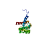
- PDB-8y6k: Cryo-EM structure of full-length MICAL1 in the autoinhibited state -
+ Open data

- Basic information
| Entry | Database: PDB / ID: 8y6k | |||||||||
|---|---|---|---|---|---|---|---|---|---|---|
| Title | Cryo-EM structure of full-length MICAL1 in the autoinhibited state | |||||||||
 Components Components | [F-actin]-monooxygenase MICAL1 | |||||||||
 Keywords Keywords | OXIDOREDUCTASE / MICAL1 / Monooxygenase / F-actin disassembly / autoinhibition | |||||||||
| Function / homology |  Function and homology information Function and homology informationhippocampal mossy fiber expansion / : / F-actin monooxygenase / F-actin monooxygenase activity / NAD(P)H oxidase (H2O2-forming) / sulfur oxidation / regulation of regulated secretory pathway / NAD(P)H oxidase H2O2-forming activity / oxidoreductase activity, acting on paired donors, with incorporation or reduction of molecular oxygen, NAD(P)H as one donor, and incorporation of one atom of oxygen / actin filament depolymerization ...hippocampal mossy fiber expansion / : / F-actin monooxygenase / F-actin monooxygenase activity / NAD(P)H oxidase (H2O2-forming) / sulfur oxidation / regulation of regulated secretory pathway / NAD(P)H oxidase H2O2-forming activity / oxidoreductase activity, acting on paired donors, with incorporation or reduction of molecular oxygen, NAD(P)H as one donor, and incorporation of one atom of oxygen / actin filament depolymerization / intermediate filament / actin filament bundle assembly / intercellular bridge / cytoskeleton organization / FAD binding / actin filament / monooxygenase activity / SH3 domain binding / small GTPase binding / actin filament binding / actin cytoskeleton / Factors involved in megakaryocyte development and platelet production / actin binding / midbody / endosome membrane / cilium / ciliary basal body / protein kinase binding / negative regulation of apoptotic process / signal transduction / nucleoplasm / metal ion binding / plasma membrane / cytosol / cytoplasm Similarity search - Function | |||||||||
| Biological species |  Homo sapiens (human) Homo sapiens (human) | |||||||||
| Method | ELECTRON MICROSCOPY / single particle reconstruction / cryo EM / Resolution: 3.94 Å | |||||||||
 Authors Authors | Niu, F. / Wei, Z. | |||||||||
| Funding support |  China, 2items China, 2items
| |||||||||
 Citation Citation | Journal: Nat Commun / Year: 2024 Title: Autoinhibition and relief mechanisms for MICAL monooxygenases in F-actin disassembly. Authors: Leishu Lin / Jiayuan Dong / Shun Xu / Jinman Xiao / Cong Yu / Fengfeng Niu / Zhiyi Wei / Abstract: MICAL proteins represent a unique family of actin regulators crucial for synapse development, membrane trafficking, and cytokinesis. Unlike classical actin regulators, MICALs catalyze the oxidation ...MICAL proteins represent a unique family of actin regulators crucial for synapse development, membrane trafficking, and cytokinesis. Unlike classical actin regulators, MICALs catalyze the oxidation of specific residues within actin filaments to induce robust filament disassembly. The potent activity of MICALs requires tight control to prevent extensive damage to actin cytoskeleton. However, the molecular mechanism governing MICALs' activity regulation remains elusive. Here, we report the cryo-EM structure of MICAL1 in the autoinhibited state, unveiling a head-to-tail interaction that allosterically blocks enzymatic activity. The structure also reveals the assembly of C-terminal domains via a tripartite interdomain interaction, stabilizing the inhibitory conformation of the RBD. Our structural, biochemical, and cellular analyses elucidate a multi-step mechanism to relieve MICAL1 autoinhibition in response to the dual-binding of two Rab effectors, revealing its intricate activity regulation mechanisms. Furthermore, our mutagenesis study of MICAL3 suggests the conserved autoinhibition and relief mechanisms among MICALs. | |||||||||
| History |
|
- Structure visualization
| Structure viewer | Molecule: MolmilJmol/JSmol |
|---|
- Downloads & links
-Download
| PDBx/mmCIF format | 8y6k.cif.gz | 151.9 KB | Display | PDBx/mmCIF format |
|---|---|---|---|---|
| PDB format | pdb8y6k.ent.gz | 111.1 KB | Display | PDB format |
| PDBx/mmJSON format | 8y6k.json.gz | Tree view | PDBx/mmJSON format | |
| Others |  Other downloads Other downloads |
-Validation report
| Summary document | 8y6k_validation.pdf.gz | 1.1 MB | Display | wwPDB validaton report |
|---|---|---|---|---|
| Full document | 8y6k_full_validation.pdf.gz | 1.1 MB | Display | |
| Data in XML | 8y6k_validation.xml.gz | 34.6 KB | Display | |
| Data in CIF | 8y6k_validation.cif.gz | 49.1 KB | Display | |
| Arichive directory | https://data.pdbj.org/pub/pdb/validation_reports/y6/8y6kftp://data.pdbj.org/pub/pdb/validation_reports/y6/8y6k | HTTPS FTP |
-Related structure data
| Related structure data |  38989MC M: map data used to model this data C: citing same article ( |
|---|---|
| Similar structure data |
-Links
 PDBj
PDBj- Assembly
| Deposited unit | 
|
|---|---|
| 1 |
|
-Components
| #1: Protein | [ Mass: 118014.734 Da / Num. of mol.: 1 Source method: isolated from a genetically manipulated source Source: (gene. exp.) Homo sapiens (human) / Gene: MICAL1, MICAL, NICAL / Cell line (production host): HEK293F / Production host: Homo sapiens (human)References: UniProt: Q8TDZ2, F-actin monooxygenase, NAD(P)H oxidase (H2O2-forming) | ||||
|---|---|---|---|---|---|
| #2: Chemical |   Mass: 65.409 Da / Num. of mol.: 2 / Source method: obtained synthetically / Formula: Zn / Feature type: SUBJECT OF INVESTIGATION Mass: 65.409 Da / Num. of mol.: 2 / Source method: obtained synthetically / Formula: Zn / Feature type: SUBJECT OF INVESTIGATION#3: Chemical | ChemComp-FAD / |   Mass: 785.550 Da / Num. of mol.: 1 / Source method: obtained synthetically / Formula: C27H33N9O15P2 / Feature type: SUBJECT OF INVESTIGATION / Comment: FAD*YM Mass: 785.550 Da / Num. of mol.: 1 / Source method: obtained synthetically / Formula: C27H33N9O15P2 / Feature type: SUBJECT OF INVESTIGATION / Comment: FAD*YMHas ligand of interest | Y | |
-Experimental details
-Experiment
| Experiment | Method: ELECTRON MICROSCOPY |
|---|---|
| EM experiment | Aggregation state: PARTICLE / 3D reconstruction method: single particle reconstruction |
- Sample preparation
| Component | Name: full-length MICAL1 protein / Type: COMPLEX / Entity ID: #1 / Source: RECOMBINANT |
|---|---|
| Molecular weight | Value: 0.106 MDa / Experimental value: YES |
| Source (natural) | Organism: Homo sapiens (human) |
| Source (recombinant) | Organism: Homo sapiens (human) / Cell: HEK293F |
| Buffer solution | pH: 7.5 Details: 50 mM Tris, pH 7.5, 100 mM NaCl, 2 mM MgCl2, 2 mM DTT. |
| Specimen | Conc.: 0.3 mg/ml / Embedding applied: NO / Shadowing applied: NO / Staining applied: NO / Vitrification applied: YES |
| Specimen support | Grid material: COPPER / Grid mesh size: 300 divisions/in. / Grid type: Quantifoil R1.2/1.3 |
| Vitrification | Instrument: FEI VITROBOT MARK IV / Cryogen name: ETHANE / Humidity: 100 % / Chamber temperature: 277.15 K |
- Electron microscopy imaging
| Experimental equipment |  Model: Titan Krios / Image courtesy: FEI Company |
|---|---|
| Microscopy | Model: FEI TITAN KRIOS |
| Electron gun | Electron source:  FIELD EMISSION GUN / Accelerating voltage: 300 kV / Illumination mode: SPOT SCAN FIELD EMISSION GUN / Accelerating voltage: 300 kV / Illumination mode: SPOT SCAN |
| Electron lens | Mode: BRIGHT FIELD / Nominal defocus max: 2500 nm / Nominal defocus min: 1500 nm |
| Image recording | Electron dose: 40 e/Å2 / Detector mode: SUPER-RESOLUTION / Film or detector model: GATAN K2 SUMMIT (4k x 4k) |
- Processing
| EM software |
| ||||||||||||||||||||||||||||||||||||||||
|---|---|---|---|---|---|---|---|---|---|---|---|---|---|---|---|---|---|---|---|---|---|---|---|---|---|---|---|---|---|---|---|---|---|---|---|---|---|---|---|---|---|
| CTF correction | Type: NONE | ||||||||||||||||||||||||||||||||||||||||
| Particle selection | Num. of particles selected: 9063162 | ||||||||||||||||||||||||||||||||||||||||
| Symmetry | Point symmetry: C1 (asymmetric) | ||||||||||||||||||||||||||||||||||||||||
| 3D reconstruction | Resolution: 3.94 Å / Resolution method: FSC 0.143 CUT-OFF / Num. of particles: 122086 / Symmetry type: POINT | ||||||||||||||||||||||||||||||||||||||||
| Atomic model building | Protocol: FLEXIBLE FIT / Space: REAL | ||||||||||||||||||||||||||||||||||||||||
| Atomic model building |
| ||||||||||||||||||||||||||||||||||||||||
| Refine LS restraints |
|