National Natural Science Foundation of China (NSFC)
31971131
China
National Natural Science Foundation of China (NSFC)
32170697
China
Citation
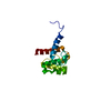
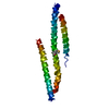
Journal: Nat Commun / Year: 2024 Title: Autoinhibition and relief mechanisms for MICAL monooxygenases in F-actin disassembly. Authors: Leishu Lin / Jiayuan Dong / Shun Xu / Jinman Xiao / Cong Yu / Fengfeng Niu / Zhiyi Wei / Abstract: MICAL proteins represent a unique family of actin regulators crucial for synapse development, membrane trafficking, and cytokinesis. Unlike classical actin regulators, MICALs catalyze the oxidation ...MICAL proteins represent a unique family of actin regulators crucial for synapse development, membrane trafficking, and cytokinesis. Unlike classical actin regulators, MICALs catalyze the oxidation of specific residues within actin filaments to induce robust filament disassembly. The potent activity of MICALs requires tight control to prevent extensive damage to actin cytoskeleton. However, the molecular mechanism governing MICALs' activity regulation remains elusive. Here, we report the cryo-EM structure of MICAL1 in the autoinhibited state, unveiling a head-to-tail interaction that allosterically blocks enzymatic activity. The structure also reveals the assembly of C-terminal domains via a tripartite interdomain interaction, stabilizing the inhibitory conformation of the RBD. Our structural, biochemical, and cellular analyses elucidate a multi-step mechanism to relieve MICAL1 autoinhibition in response to the dual-binding of two Rab effectors, revealing its intricate activity regulation mechanisms. Furthermore, our mutagenesis study of MICAL3 suggests the conserved autoinhibition and relief mechanisms among MICALs.
In the structure databanks used in Yorodumi, some data are registered as the other names, "COVID-19 virus" and "2019-nCoV". Here are the details of the virus and the list of structure data.
Jan 31, 2019. EMDB accession codes are about to change! (news from PDBe EMDB page)
EMDB accession codes are about to change! (news from PDBe EMDB page)
The allocation of 4 digits for EMDB accession codes will soon come to an end. Whilst these codes will remain in use, new EMDB accession codes will include an additional digit and will expand incrementally as the available range of codes is exhausted. The current 4-digit format prefixed with “EMD-” (i.e. EMD-XXXX) will advance to a 5-digit format (i.e. EMD-XXXXX), and so on. It is currently estimated that the 4-digit codes will be depleted around Spring 2019, at which point the 5-digit format will come into force.
The EM Navigator/Yorodumi systems omit the EMD- prefix.
Related info.:Q: What is EMD? / ID/Accession-code notation in Yorodumi/EM Navigator
Yorodumi is a browser for structure data from EMDB, PDB, SASBDB, etc.
This page is also the successor to EM Navigator detail page, and also detail information page/front-end page for Omokage search.
The word "yorodu" (or yorozu) is an old Japanese word meaning "ten thousand". "mi" (miru) is to see.
Related info.:EMDB / PDB / SASBDB / Comparison of 3 databanks / Yorodumi Search / Aug 31, 2016. New EM Navigator & Yorodumi / Yorodumi Papers / Jmol/JSmol / Function and homology information / Changes in new EM Navigator and Yorodumi
 Movie
Movie Controller

 Yorodumi
 Open data
 Basic information
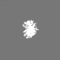
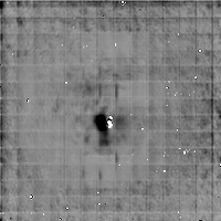
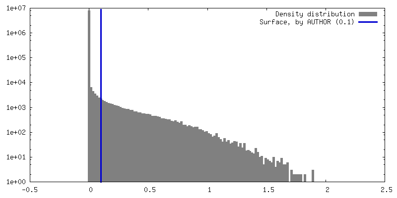
 Map data
 Sample
 Keywords
 Function and homology information
 Homo sapiens (human)
 Authors
 China, 2 items
  Citation
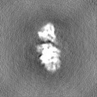
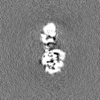
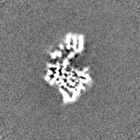
 Structure visualization
 Downloads & links
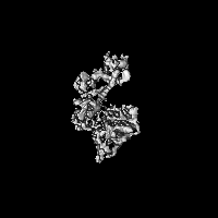
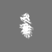
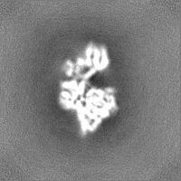
 emd_38989.png
 http://ftp.pdbj.org/pub/emdb/structures/EMD-38989
 Z (Sec.)
 Y (Row.)
 X (Col.)


 Sample components
 Processing
 Electron microscopy
 FIELD EMISSION GUN