 Movie
Movie Controller

[English] 日本語
 Yorodumi
- PDB-8y6h: P-glycoprotein in complex with UIC2 Fab and triple elacridar mole... -
+ Open data

- Basic information
| Entry | Database: PDB / ID: 8y6h | ||||||||||||
|---|---|---|---|---|---|---|---|---|---|---|---|---|---|
| Title | P-glycoprotein in complex with UIC2 Fab and triple elacridar molecules in LMNG detergent | ||||||||||||
 Components Components |
| ||||||||||||
 Keywords Keywords | MEMBRANE PROTEIN / ABC transporter / elacridar / P-glycoprotein | ||||||||||||
| Function / homology |  Function and homology information Function and homology informationhormone transport / phosphatidylethanolamine floppase activity / cellular response to nonylphenol / cellular response to borneol / response to codeine / cellular response to mycotoxin / daunorubicin transport / positive regulation of response to drug / terpenoid transport / ceramide floppase activity ...hormone transport / phosphatidylethanolamine floppase activity / cellular response to nonylphenol / cellular response to borneol / response to codeine / cellular response to mycotoxin / daunorubicin transport / positive regulation of response to drug / terpenoid transport / ceramide floppase activity / regulation of intestinal absorption / cellular response to external biotic stimulus / response to cyclosporin A / response to antineoplastic agent / positive regulation of establishment of Sertoli cell barrier / negative regulation of sensory perception of pain / carboxylic acid transmembrane transport / floppase activity / ceramide translocation / Abacavir transmembrane transport / response to quercetin / carboxylic acid transmembrane transporter activity / establishment of blood-retinal barrier / phosphatidylethanolamine flippase activity / protein localization to bicellular tight junction / phosphatidylcholine floppase activity / external side of apical plasma membrane / Atorvastatin ADME / response to thyroxine / xenobiotic transport across blood-brain barrier / establishment of blood-brain barrier / export across plasma membrane / P-type phospholipid transporter / xenobiotic detoxification by transmembrane export across the plasma membrane / transepithelial transport / cellular response to L-glutamate / response to vitamin A / ABC-type xenobiotic transporter / response to glucagon / response to vitamin D / intestinal absorption / response to alcohol / response to glycoside / Prednisone ADME / phospholipid translocation / ABC-type xenobiotic transporter activity / cellular hyperosmotic salinity response / cellular response to alkaloid / cellular response to antibiotic / maintenance of blood-brain barrier / ATPase-coupled transmembrane transporter activity / efflux transmembrane transporter activity / xenobiotic transmembrane transporter activity / cellular response to dexamethasone stimulus / response to cadmium ion / transmembrane transporter activity / transport across blood-brain barrier / lactation / response to progesterone / xenobiotic metabolic process / regulation of chloride transport / placenta development / stem cell proliferation / bioluminescence / cellular response to estradiol stimulus / brush border membrane / female pregnancy / circadian rhythm / transmembrane transport / G2/M transition of mitotic cell cycle / cellular response to tumor necrosis factor / ABC-family protein mediated transport / cellular response to lipopolysaccharide / response to hypoxia / apical plasma membrane / response to xenobiotic stimulus / ubiquitin protein ligase binding / cell surface / ATP hydrolysis activity / extracellular exosome / ATP binding / membrane / plasma membrane / cytoplasm Similarity search - Function | ||||||||||||
| Biological species |  Homo sapiens (human) Homo sapiens (human) | ||||||||||||
| Method | ELECTRON MICROSCOPY / single particle reconstruction / cryo EM / Resolution: 2.49 Å | ||||||||||||
 Authors Authors | Hamaguchi-Suzuki, N. / Adachi, N. / Moriya, T. / Kawasaki, M. / Suzuki, K. / Anzai, N. / Senda, T. / Murata, T. | ||||||||||||
| Funding support |  Japan, 3items Japan, 3items
| ||||||||||||
 Citation Citation | Journal: Biochem Biophys Res Commun / Year: 2024 Title: Cryo-EM structure of P-glycoprotein bound to triple elacridar inhibitor molecules. Authors: Norie Hamaguchi-Suzuki / Naruhiko Adachi / Toshio Moriya / Satoshi Yasuda / Masato Kawasaki / Kano Suzuki / Satoshi Ogasawara / Naohiko Anzai / Toshiya Senda / Takeshi Murata / Abstract: P-glycoprotein (P-gp) is an ATP-binding cassette transporter known for its roles in expelling xenobiotic compounds from cells and contributing to cellular drug resistance through multidrug efflux. ...P-glycoprotein (P-gp) is an ATP-binding cassette transporter known for its roles in expelling xenobiotic compounds from cells and contributing to cellular drug resistance through multidrug efflux. This mechanism is particularly problematic in cancer cells, where it diminishes the therapeutic efficacy of anticancer drugs. P-gp inhibitors, such as elacridar, have been developed to circumvent the decrease in drug efficacy due to P-gp efflux. An earlier study reported the cryo-EM structure of human P-gp-Fab (MRK-16) complex bound by two elacridar molecules, at a resolution of 3.6 Å. In this study, we have obtained a higher resolution (2.5 Å) structure of the P-gp- Fab (UIC2) complex bound by three elacridar molecules. This finding, which exposes a larger space for compound-binding sites than previously acknowledged, has significant implications for the development of more selective inhibitors and enhances our understanding of the compound recognition mechanism of P-gp. | ||||||||||||
| History |
|
- Structure visualization
| Structure viewer | Molecule: MolmilJmol/JSmol |
|---|
- Downloads & links
-Download
| PDBx/mmCIF format | 8y6h.cif.gz | 236.8 KB | Display | PDBx/mmCIF format |
|---|---|---|---|---|
| PDB format | pdb8y6h.ent.gz | 146.2 KB | Display | PDB format |
| PDBx/mmJSON format | 8y6h.json.gz | Tree view | PDBx/mmJSON format | |
| Others |  Other downloads Other downloads |
-Validation report
| Arichive directory | https://data.pdbj.org/pub/pdb/validation_reports/y6/8y6hftp://data.pdbj.org/pub/pdb/validation_reports/y6/8y6h | HTTPS FTP |
|---|
-Related structure data
| Related structure data |  38986MC 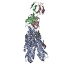 8y6iC M: map data used to model this data C: citing same article ( |
|---|---|
| Similar structure data |

-Links
 PDBj
PDBj


- Assembly
| Deposited unit | 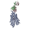
|
|---|---|
| 1 |
|
-Components
| #1: Protein | Mass: 170535.812 Da / Num. of mol.: 1 Source method: isolated from a genetically manipulated source Source: (gene. exp.) Homo sapiens (human) / Gene: ABCB1, MDR1, PGY1, blFP-Y3 / Production host: Homo sapiens (human)References: UniProt: P08183, UniProt: A0A1S4NYF2, ABC-type xenobiotic transporter, P-type phospholipid transporter | ||||
|---|---|---|---|---|---|
| #2: Antibody | Mass: 24321.039 Da / Num. of mol.: 1 Source method: isolated from a genetically manipulated source Source: (gene. exp.) | ||||
| #3: Antibody | Mass: 24381.281 Da / Num. of mol.: 1 Source method: isolated from a genetically manipulated source Source: (gene. exp.) | ||||
| #4: Chemical |   Mass: 563.643 Da / Num. of mol.: 3 / Source method: obtained synthetically / Formula: C34H33N3O5 / Feature type: SUBJECT OF INVESTIGATION Mass: 563.643 Da / Num. of mol.: 3 / Source method: obtained synthetically / Formula: C34H33N3O5 / Feature type: SUBJECT OF INVESTIGATIONHas ligand of interest | Y | Has protein modification | Y | |
-Experimental details
-Experiment
| Experiment | Method: ELECTRON MICROSCOPY |
|---|---|
| EM experiment | Aggregation state: PARTICLE / 3D reconstruction method: single particle reconstruction |
- Sample preparation
| Component | Name: Multidrug resistance protein / Type: COMPLEX / Entity ID: #1-#3 / Source: RECOMBINANT |
|---|---|
| Source (natural) | Organism: Homo sapiens (human) |
| Source (recombinant) | Organism: Homo sapiens (human) |
| Buffer solution | pH: 7.5 |
| Specimen | Embedding applied: NO / Shadowing applied: NO / Staining applied: NO / Vitrification applied: YES |
| Vitrification | Instrument: FEI VITROBOT MARK IV / Cryogen name: ETHANE / Humidity: 100 % / Chamber temperature: 291 K |
- Electron microscopy imaging
| Experimental equipment |  Model: Titan Krios / Image courtesy: FEI Company |
|---|---|
| Microscopy | Model: TFS KRIOS |
| Electron gun | Electron source:  FIELD EMISSION GUN / Accelerating voltage: 300 kV / Illumination mode: FLOOD BEAM FIELD EMISSION GUN / Accelerating voltage: 300 kV / Illumination mode: FLOOD BEAM |
| Electron lens | Mode: BRIGHT FIELD / Nominal defocus max: 20000 nm / Nominal defocus min: 800 nm / Cs: 2.7 mm |
| Image recording | Electron dose: 48.8 e/Å2 / Film or detector model: GATAN K3 BIOQUANTUM (6k x 4k) / Num. of grids imaged: 1 / Num. of real images: 4482 |
- Processing
| EM software |
| ||||||||||||||||||||||||
|---|---|---|---|---|---|---|---|---|---|---|---|---|---|---|---|---|---|---|---|---|---|---|---|---|---|
| CTF correction | Type: PHASE FLIPPING AND AMPLITUDE CORRECTION | ||||||||||||||||||||||||
| 3D reconstruction | Resolution: 2.49 Å / Resolution method: FSC 0.143 CUT-OFF / Num. of particles: 142821 / Symmetry type: POINT | ||||||||||||||||||||||||
| Atomic model building | Space: RECIPROCAL | ||||||||||||||||||||||||
| Atomic model building | PDB-ID: 6QEX Accession code: 6QEX / Source name: PDB / Type: experimental model | ||||||||||||||||||||||||
| Refinement | Cross valid method: NONE Stereochemistry target values: GeoStd + Monomer Library + CDL v1.2 | ||||||||||||||||||||||||
| Displacement parameters | Biso mean: 110.82 Å2 | ||||||||||||||||||||||||
| Refine LS restraints |
|

