 Movie
Movie Controller

[English] 日本語
 Yorodumi
- PDB-8y4b: Crystal structure of L-2-keto-3-deoxyfuconate 4-dehydrogenase bou... -
+ Open data

- Basic information
| Entry | Database: PDB / ID: 8y4b | ||||||
|---|---|---|---|---|---|---|---|
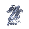
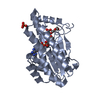
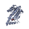
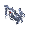
| Title | Crystal structure of L-2-keto-3-deoxyfuconate 4-dehydrogenase bound to L-2,4-DKDF and NADH | ||||||
 Components Components | SDR family oxidoreductase | ||||||
 Keywords Keywords | OXIDOREDUCTASE / dehydrogenase / Rossmann Fold | ||||||
| Function / homology | : / NICOTINAMIDE-ADENINE-DINUCLEOTIDE / :  Function and homology information Function and homology information | ||||||
| Biological species |  Herbaspirillum huttiense (bacteria) Herbaspirillum huttiense (bacteria) | ||||||
| Method |  X-RAY DIFFRACTION / SYNCHROTRON / MOLECULAR REPLACEMENT / Resolution: 1.67 Å X-RAY DIFFRACTION / SYNCHROTRON / MOLECULAR REPLACEMENT / Resolution: 1.67 Å | ||||||
 Authors Authors | Akagashi, M. / Watanabe, S. | ||||||
| Funding support | 1items
| ||||||
 Citation Citation | Journal: Sci Rep / Year: 2024 Title: Crystal structure of L-2-keto-3-deoxyfuconate 4-dehydrogenase reveals a unique binding mode as a alpha-furanosyl hemiketal of substrates. Authors: Akagashi, M. / Watanabe, S. / Kwiatkowski, S. / Drozak, J. / Terawaki, S. / Watanabe, Y. | ||||||
| History |
|
- Structure visualization
| Structure viewer | Molecule: MolmilJmol/JSmol |
|---|
- Downloads & links
-Download
| PDBx/mmCIF format | 8y4b.cif.gz | 200.9 KB | Display | PDBx/mmCIF format |
|---|---|---|---|---|
| PDB format | pdb8y4b.ent.gz | Display | PDB format | |
| PDBx/mmJSON format | 8y4b.json.gz | Tree view | PDBx/mmJSON format | |
| Others |  Other downloads Other downloads |
-Validation report
| Summary document | 8y4b_validation.pdf.gz | 2 MB | Display | wwPDB validaton report |
|---|---|---|---|---|
| Full document | 8y4b_full_validation.pdf.gz | 2 MB | Display | |
| Data in XML | 8y4b_validation.xml.gz | 42.4 KB | Display | |
| Data in CIF | 8y4b_validation.cif.gz | 58 KB | Display | |
| Arichive directory | https://data.pdbj.org/pub/pdb/validation_reports/y4/8y4bftp://data.pdbj.org/pub/pdb/validation_reports/y4/8y4b | HTTPS FTP |
-Related structure data
-Links
 PDBj
PDBj- Assembly
| Deposited unit | 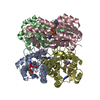
| ||||||||
|---|---|---|---|---|---|---|---|---|---|
| 1 | 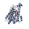
| ||||||||
| 2 | 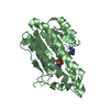
| ||||||||
| 3 | 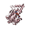
| ||||||||
| 4 | 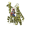
| ||||||||
| Unit cell |
|
-Components
| #1: Protein | Mass: 27596.275 Da / Num. of mol.: 4 Source method: isolated from a genetically manipulated source Source: (gene. exp.) Herbaspirillum huttiense (bacteria) / Gene: E2K99_19890 / Production host: #2: Chemical | ChemComp-NAD /   Mass: 663.425 Da / Num. of mol.: 4 / Source method: obtained synthetically / Formula: C21H27N7O14P2 / Feature type: SUBJECT OF INVESTIGATION / Comment: NAD*YM Mass: 663.425 Da / Num. of mol.: 4 / Source method: obtained synthetically / Formula: C21H27N7O14P2 / Feature type: SUBJECT OF INVESTIGATION / Comment: NAD*YM#3: Chemical | ChemComp-A1LXB / | Mass: 160.125 Da / Num. of mol.: 1 / Source method: obtained synthetically / Formula: C6H8O5 / Feature type: SUBJECT OF INVESTIGATION #4: Water | ChemComp-HOH / |  Mass: 18.015 Da / Num. of mol.: 393 / Source method: isolated from a natural source / Formula: H2O Mass: 18.015 Da / Num. of mol.: 393 / Source method: isolated from a natural source / Formula: H2OHas ligand of interest | Y | |
|---|
-Experimental details
-Experiment
| Experiment | Method: X-RAY DIFFRACTION / Number of used crystals: 1 |
|---|
- Sample preparation
| Crystal | Density Matthews: 2.28 Å3/Da / Density % sol: 45.96 % |
|---|---|
| Crystal grow | Temperature: 293 K / Method: evaporation Details: 0.2 M Sodium acetate, 0.1 M Tris-HCl pH8.5, 25% PEG4000 |
-Data collection
| Diffraction | Mean temperature: 293 K / Serial crystal experiment: N |
|---|---|
| Diffraction source | Source: SYNCHROTRON / Site: SPring-8  / Beamline: BL45XU / Wavelength: 1 Å / Beamline: BL45XU / Wavelength: 1 Å |
| Detector | Type: DECTRIS PILATUS3 6M / Detector: PIXEL / Date: Oct 27, 2023 |
| Radiation | Protocol: SINGLE WAVELENGTH / Monochromatic (M) / Laue (L): M / Scattering type: x-ray |
| Radiation wavelength | Wavelength: 1 Å / Relative weight: 1 |
| Reflection | Resolution: 1.67→44.32 Å / Num. obs: 110122 / % possible obs: 100 % / Redundancy: 19.57 % / CC1/2: 0.991 / Net I/σ(I): 10.1 |
| Reflection shell | Resolution: 1.67→1.7 Å / Num. unique obs: 5406 / CC1/2: 0.759 |
- Processing
| Software |
| |||||||||||||||||||||||||||||||||||||||||||||||||||||||||||||||||||||||||||||||||||||||||||||||||||||||||||||||||||||||||||||||||||||||||||||||||||||||||||||||||||||||||||||||||||||||||||||||||||||||||||||||||||||||||
|---|---|---|---|---|---|---|---|---|---|---|---|---|---|---|---|---|---|---|---|---|---|---|---|---|---|---|---|---|---|---|---|---|---|---|---|---|---|---|---|---|---|---|---|---|---|---|---|---|---|---|---|---|---|---|---|---|---|---|---|---|---|---|---|---|---|---|---|---|---|---|---|---|---|---|---|---|---|---|---|---|---|---|---|---|---|---|---|---|---|---|---|---|---|---|---|---|---|---|---|---|---|---|---|---|---|---|---|---|---|---|---|---|---|---|---|---|---|---|---|---|---|---|---|---|---|---|---|---|---|---|---|---|---|---|---|---|---|---|---|---|---|---|---|---|---|---|---|---|---|---|---|---|---|---|---|---|---|---|---|---|---|---|---|---|---|---|---|---|---|---|---|---|---|---|---|---|---|---|---|---|---|---|---|---|---|---|---|---|---|---|---|---|---|---|---|---|---|---|---|---|---|---|---|---|---|---|---|---|---|---|---|---|---|---|---|---|---|---|
| Refinement | Method to determine structure: MOLECULAR REPLACEMENT Starting model: AlphaFold Resolution: 1.67→44.32 Å / SU ML: 0.32 / Cross valid method: NONE / σ(F): 1.33 / Phase error: 34.94 / Stereochemistry target values: ML
| |||||||||||||||||||||||||||||||||||||||||||||||||||||||||||||||||||||||||||||||||||||||||||||||||||||||||||||||||||||||||||||||||||||||||||||||||||||||||||||||||||||||||||||||||||||||||||||||||||||||||||||||||||||||||
| Solvent computation | Shrinkage radii: 0.9 Å / VDW probe radii: 1.1 Å / Solvent model: FLAT BULK SOLVENT MODEL | |||||||||||||||||||||||||||||||||||||||||||||||||||||||||||||||||||||||||||||||||||||||||||||||||||||||||||||||||||||||||||||||||||||||||||||||||||||||||||||||||||||||||||||||||||||||||||||||||||||||||||||||||||||||||
| Refinement step | Cycle: LAST / Resolution: 1.67→44.32 Å
| |||||||||||||||||||||||||||||||||||||||||||||||||||||||||||||||||||||||||||||||||||||||||||||||||||||||||||||||||||||||||||||||||||||||||||||||||||||||||||||||||||||||||||||||||||||||||||||||||||||||||||||||||||||||||
| Refine LS restraints |
| |||||||||||||||||||||||||||||||||||||||||||||||||||||||||||||||||||||||||||||||||||||||||||||||||||||||||||||||||||||||||||||||||||||||||||||||||||||||||||||||||||||||||||||||||||||||||||||||||||||||||||||||||||||||||
| LS refinement shell |
|
