 Movie
Movie Controller


+ Open data

- Basic information
| Entry | Database: PDB / ID: 8xwd | ||||||||||||||||||||||||
|---|---|---|---|---|---|---|---|---|---|---|---|---|---|---|---|---|---|---|---|---|---|---|---|---|---|
| Title | Croy-EM structure of alpha synuclein fibril with EGCG | ||||||||||||||||||||||||
 Components Components | Alpha-synuclein | ||||||||||||||||||||||||
 Keywords Keywords | PROTEIN FIBRIL / amyloid | ||||||||||||||||||||||||
| Function / homology |  Function and homology information Function and homology informationnegative regulation of mitochondrial electron transport, NADH to ubiquinone / response to desipramine / negative regulation of dopamine uptake involved in synaptic transmission / negative regulation of norepinephrine uptake / positive regulation of SNARE complex assembly / positive regulation of hydrogen peroxide catabolic process / supramolecular fiber / negative regulation of chaperone-mediated autophagy / regulation of synaptic vesicle recycling / regulation of reactive oxygen species biosynthetic process ...negative regulation of mitochondrial electron transport, NADH to ubiquinone / response to desipramine / negative regulation of dopamine uptake involved in synaptic transmission / negative regulation of norepinephrine uptake / positive regulation of SNARE complex assembly / positive regulation of hydrogen peroxide catabolic process / supramolecular fiber / negative regulation of chaperone-mediated autophagy / regulation of synaptic vesicle recycling / regulation of reactive oxygen species biosynthetic process / positive regulation of protein localization to cell periphery / negative regulation of exocytosis / dopamine biosynthetic process / dopamine uptake involved in synaptic transmission / negative regulation of dopamine metabolic process / response to iron(II) ion / negative regulation of platelet-derived growth factor receptor signaling pathway / SNARE complex assembly / negative regulation of microtubule polymerization / negative regulation of thrombin-activated receptor signaling pathway / synaptic vesicle priming / Lewy body / synaptic vesicle transport / regulation of norepinephrine uptake / synaptic vesicle exocytosis / transporter regulator activity / protein kinase inhibitor activity / positive regulation of inositol phosphate biosynthetic process / positive regulation of receptor recycling / cuprous ion binding / positive regulation of exocytosis / nuclear outer membrane / dynein complex binding / synaptic transmission, dopaminergic / regulation of dopamine secretion / positive regulation of endocytosis / response to magnesium ion / negative regulation of serotonin uptake / kinesin binding / cysteine-type endopeptidase inhibitor activity / regulation of presynapse assembly / synaptic vesicle endocytosis / alpha-tubulin binding / beta-tubulin binding / phospholipase binding / behavioral response to cocaine / cellular response to fibroblast growth factor stimulus / supramolecular fiber organization / response to type II interferon / cellular response to epinephrine stimulus / inclusion body / response to interleukin-1 / Hsp70 protein binding / axon terminus / cellular response to copper ion / positive regulation of release of sequestered calcium ion into cytosol / enzyme inhibitor activity / glutathione metabolic process / SNARE binding / protein tetramerization / regulation of microtubule cytoskeleton organization / phosphoprotein binding / receptor internalization / protein destabilization / microglial cell activation / tubulin binding / ferrous iron binding / protein sequestering activity / phospholipid binding / synapse organization / PKR-mediated signaling / tau protein binding / enzyme activator activity / positive regulation of inflammatory response / actin cytoskeleton / terminal bouton / synaptic vesicle membrane / negative regulation of neuron apoptotic process / histone binding / actin binding / response to lipopolysaccharide / growth cone / cellular response to oxidative stress / cell cortex / oxidoreductase activity / microtubule binding / amyloid fibril formation / mitochondrial outer membrane / lysosome / transcription cis-regulatory region binding / mitochondrial inner membrane / positive regulation of apoptotic process / ribosome / mitochondrial matrix / Amyloid fiber formation / copper ion binding / protein domain specific binding / axon / lipid binding / neuronal cell body Similarity search - Function | ||||||||||||||||||||||||
| Biological species |  Homo sapiens (human) Homo sapiens (human) | ||||||||||||||||||||||||
| Method | ELECTRON MICROSCOPY / helical reconstruction / cryo EM / Resolution: 3.1 Å | ||||||||||||||||||||||||
 Authors Authors | Li, X. / Liu, C. | ||||||||||||||||||||||||
| Funding support | 1items
| ||||||||||||||||||||||||
 Citation Citation | Journal: Adv Sci (Weinh) / Year: 2025 Title: Single-Molecule Insight Into α-Synuclein Fibril Structure and Mechanics Modulated by Chemical Compounds. Authors: Xiang Li / Lulu Bi / Shenqing Zhang / Qianhui Xu / Wencheng Xia / Youqi Tao / Shaojuan Wu / Yanan Li / Weidong Le / Wenyan Kang / Dan Li / Bo Sun / Cong Liu /  Abstract: α-Syn fibrils, a key pathological hallmark of Parkinson's disease, is closely associated with disease initiation and progression. Several small molecules are found to bind or dissolve α-syn ...α-Syn fibrils, a key pathological hallmark of Parkinson's disease, is closely associated with disease initiation and progression. Several small molecules are found to bind or dissolve α-syn fibrils, offering potential therapeutic applications. Here, an innovative optical tweezers-based, fluorescence-combined approach is developed to probe the mechanical characteristics of α-syn fibrils at the single-molecule level. When subjected to axial stretching, local deformation within α-syn fibrils appeared at forces above 50 pN. These structural alternations occurred stepwise and are irreversible, suggesting unfolding of individual α-syn molecules or subdomains. Additionally, α-syn fibrils exhibits high heterogeneity in lateral disruption, with rupture force ranging from 50 to 500 pN. The impact of different compounds on the structure and mechanical features of α-syn fibrils is further examined. Notably, epigallocatechin gallate (EGCG) generally attenuates the rupture force of fibrils by wedging into the N-terminal polar groove and induces fibril dissociation. Conversely, copper chlorophyllin A (CCA) attaches to four different sites wrapping around the fibril core, reinforcing the stability of the fibril against rupture forces. The work offers an effective method for characterizing single-fibril properties and bridges compound-induced structural alternations with mechanical response. These insights are valuable for understanding amyloid fibril mechanics and their regulation by small molecules. | ||||||||||||||||||||||||
| History |
|
- Structure visualization
| Structure viewer | Molecule: MolmilJmol/JSmol |
|---|
- Downloads & links
-Download
| PDBx/mmCIF format | 8xwd.cif.gz | 73.7 KB | Display | PDBx/mmCIF format |
|---|---|---|---|---|
| PDB format | pdb8xwd.ent.gz | 54.3 KB | Display | PDB format |
| PDBx/mmJSON format | 8xwd.json.gz | Tree view | PDBx/mmJSON format | |
| Others |  Other downloads Other downloads |
-Validation report
| Arichive directory | https://data.pdbj.org/pub/pdb/validation_reports/xw/8xwdftp://data.pdbj.org/pub/pdb/validation_reports/xw/8xwd | HTTPS FTP |
|---|
-Related structure data
| Related structure data |  38733MC M: map data used to model this data C: citing same article ( |
|---|---|
| Similar structure data |
-Links
 PDBj
PDBj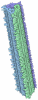
- Assembly
| Deposited unit | 
|
|---|---|
| 1 |
|
-Components
| #1: Protein | Mass: 14476.108 Da / Num. of mol.: 6 Source method: isolated from a genetically manipulated source Details: EGCG / Source: (gene. exp.) Homo sapiens (human) / Gene: SNCA, NACP, PARK1 / Production host:   Escherichia coli K-12 (bacteria) / References: UniProt: P37840 Escherichia coli K-12 (bacteria) / References: UniProt: P37840#2: Chemical | ChemComp-KDH / ( 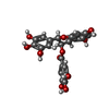 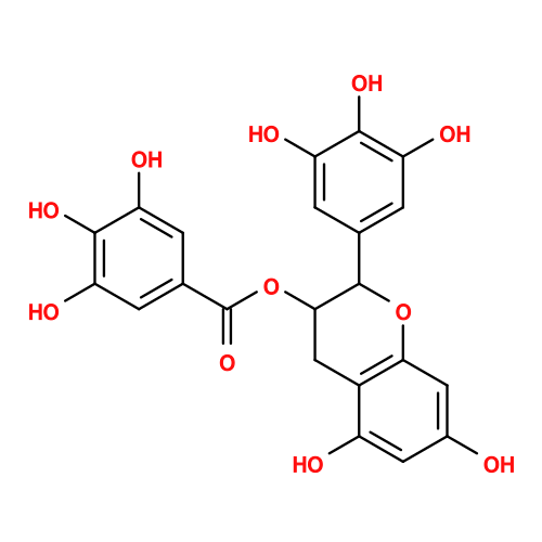 Mass: 458.372 Da / Num. of mol.: 6 / Source method: obtained synthetically / Formula: C22H18O11 / Feature type: SUBJECT OF INVESTIGATION Mass: 458.372 Da / Num. of mol.: 6 / Source method: obtained synthetically / Formula: C22H18O11 / Feature type: SUBJECT OF INVESTIGATIONHas ligand of interest | Y | Has protein modification | N | |
|---|
-Experimental details
-Experiment
| Experiment | Method: ELECTRON MICROSCOPY |
|---|---|
| EM experiment | Aggregation state: FILAMENT / 3D reconstruction method: helical reconstruction |
- Sample preparation
| Component | Name: Croy-EM structure of alpha synuclein fibril with EGCG / Type: ORGANELLE OR CELLULAR COMPONENT / Entity ID: #1 / Source: RECOMBINANT |
|---|---|
| Molecular weight | Experimental value: NO |
| Source (natural) | Organism: Homo sapiens (human) |
| Source (recombinant) | Organism: Escherichia coli K-12 (bacteria) |
| Buffer solution | pH: 7.4 |
| Specimen | Embedding applied: NO / Shadowing applied: NO / Staining applied: NO / Vitrification applied: YES |
| Vitrification | Cryogen name: ETHANE |
- Electron microscopy imaging
| Experimental equipment |  Model: Titan Krios / Image courtesy: FEI Company |
|---|---|
| Microscopy | Model: FEI TITAN KRIOS |
| Electron gun | Electron source:  FIELD EMISSION GUN / Accelerating voltage: 300 kV / Illumination mode: FLOOD BEAM FIELD EMISSION GUN / Accelerating voltage: 300 kV / Illumination mode: FLOOD BEAM |
| Electron lens | Mode: BRIGHT FIELD / Nominal defocus max: 2200 nm / Nominal defocus min: 1400 nm |
| Image recording | Electron dose: 55 e/Å2 / Film or detector model: GATAN K3 (6k x 4k) |
- Processing
| CTF correction | Type: PHASE FLIPPING AND AMPLITUDE CORRECTION |
|---|---|
| Helical symmerty | Angular rotation/subunit: -0.746 ° / Axial rise/subunit: 4.82 Å / Axial symmetry: C1 |
| 3D reconstruction | Resolution: 3.1 Å / Resolution method: FSC 0.143 CUT-OFF / Num. of particles: 207014 / Symmetry type: HELICAL |
