Journal: Adv Sci (Weinh) / Year: 2025 Title: Single-Molecule Insight Into α-Synuclein Fibril Structure and Mechanics Modulated by Chemical Compounds. Authors: Xiang Li / Lulu Bi / Shenqing Zhang / Qianhui Xu / Wencheng Xia / Youqi Tao / Shaojuan Wu / Yanan Li / Weidong Le / Wenyan Kang / Dan Li / Bo Sun / Cong Liu / Abstract: α-Syn fibrils, a key pathological hallmark of Parkinson's disease, is closely associated with disease initiation and progression. Several small molecules are found to bind or dissolve α-syn ...α-Syn fibrils, a key pathological hallmark of Parkinson's disease, is closely associated with disease initiation and progression. Several small molecules are found to bind or dissolve α-syn fibrils, offering potential therapeutic applications. Here, an innovative optical tweezers-based, fluorescence-combined approach is developed to probe the mechanical characteristics of α-syn fibrils at the single-molecule level. When subjected to axial stretching, local deformation within α-syn fibrils appeared at forces above 50 pN. These structural alternations occurred stepwise and are irreversible, suggesting unfolding of individual α-syn molecules or subdomains. Additionally, α-syn fibrils exhibits high heterogeneity in lateral disruption, with rupture force ranging from 50 to 500 pN. The impact of different compounds on the structure and mechanical features of α-syn fibrils is further examined. Notably, epigallocatechin gallate (EGCG) generally attenuates the rupture force of fibrils by wedging into the N-terminal polar groove and induces fibril dissociation. Conversely, copper chlorophyllin A (CCA) attaches to four different sites wrapping around the fibril core, reinforcing the stability of the fibril against rupture forces. The work offers an effective method for characterizing single-fibril properties and bridges compound-induced structural alternations with mechanical response. These insights are valuable for understanding amyloid fibril mechanics and their regulation by small molecules.
In the structure databanks used in Yorodumi, some data are registered as the other names, "COVID-19 virus" and "2019-nCoV". Here are the details of the virus and the list of structure data.
Jan 31, 2019. EMDB accession codes are about to change! (news from PDBe EMDB page)
EMDB accession codes are about to change! (news from PDBe EMDB page)
The allocation of 4 digits for EMDB accession codes will soon come to an end. Whilst these codes will remain in use, new EMDB accession codes will include an additional digit and will expand incrementally as the available range of codes is exhausted. The current 4-digit format prefixed with “EMD-” (i.e. EMD-XXXX) will advance to a 5-digit format (i.e. EMD-XXXXX), and so on. It is currently estimated that the 4-digit codes will be depleted around Spring 2019, at which point the 5-digit format will come into force.
The EM Navigator/Yorodumi systems omit the EMD- prefix.
Related info.:Q: What is EMD? / ID/Accession-code notation in Yorodumi/EM Navigator
Yorodumi is a browser for structure data from EMDB, PDB, SASBDB, etc.
This page is also the successor to EM Navigator detail page, and also detail information page/front-end page for Omokage search.
The word "yorodu" (or yorozu) is an old Japanese word meaning "ten thousand". "mi" (miru) is to see.
Related info.:EMDB / PDB / SASBDB / Comparison of 3 databanks / Yorodumi Search / Aug 31, 2016. New EM Navigator & Yorodumi / Yorodumi Papers / Jmol/JSmol / Function and homology information / Changes in new EM Navigator and Yorodumi
 Movie
Movie Controller


 Open data
 Basic information
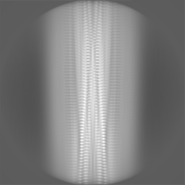
 Map data
 Sample
 Keywords
 Function and homology information
 Homo sapiens (human)
 Authors
 Citation
 Structure visualization
 Downloads & links
 emd_38733.png
 http://ftp.pdbj.org/pub/emdb/structures/EMD-38733
 Z (Sec.)
 Y (Row.)
 X (Col.)


 Sample components
 Processing
 Electron microscopy
 FIELD EMISSION GUN

