 Movie
Movie Controller

[English] 日本語
 Yorodumi
- PDB-8xvq: Crystal structure of inulosucrase from Lactobacillus reuteri 121 ... -
+ Open data

- Basic information
| Entry | Database: PDB / ID: 8xvq | ||||||||||||
|---|---|---|---|---|---|---|---|---|---|---|---|---|---|
| Title | Crystal structure of inulosucrase from Lactobacillus reuteri 121 in complex with fructose | ||||||||||||
 Components Components | Inulosucrase | ||||||||||||
 Keywords Keywords | HYDROLASE / Glycoside hydrolase 68 enzyme | ||||||||||||
| Function / homology |  Function and homology information Function and homology informationinulosucrase / inulosucrase activity / levansucrase activity / carbohydrate utilization / metal ion binding Similarity search - Function | ||||||||||||
| Biological species |  Limosilactobacillus reuteri (bacteria) Limosilactobacillus reuteri (bacteria) | ||||||||||||
| Method |  X-RAY DIFFRACTION / SYNCHROTRON / MOLECULAR REPLACEMENT / Resolution: 2.14 Å X-RAY DIFFRACTION / SYNCHROTRON / MOLECULAR REPLACEMENT / Resolution: 2.14 Å | ||||||||||||
 Authors Authors | Ni, D. / Hou, X. / Cheng, M. / Xu, W. / Rao, Y. / Mu, W. | ||||||||||||
| Funding support |  China, 3items China, 3items
| ||||||||||||
 Citation Citation | Journal: J.Agric.Food Chem. / Year: 2025 Title: Structure-Guided Tunnel Engineering to Reveal the Molecular Basis of Sugar Chain Extension of Inulosucrase. Authors: Ni, D. / Huang, Z. / Zhang, S. / Hou, X. / Xu, W. / Zhang, W. / Rao, Y. / Mu, W. | ||||||||||||
| History |
|
- Structure visualization
| Structure viewer | Molecule: MolmilJmol/JSmol |
|---|
- Downloads & links
-Download
| PDBx/mmCIF format | 8xvq.cif.gz | 227.9 KB | Display | PDBx/mmCIF format |
|---|---|---|---|---|
| PDB format | pdb8xvq.ent.gz | 174.8 KB | Display | PDB format |
| PDBx/mmJSON format | 8xvq.json.gz | Tree view | PDBx/mmJSON format | |
| Others |  Other downloads Other downloads |
-Validation report
| Arichive directory | https://data.pdbj.org/pub/pdb/validation_reports/xv/8xvqftp://data.pdbj.org/pub/pdb/validation_reports/xv/8xvq | HTTPS FTP |
|---|
-Related structure data


-Links
 PDBj
PDBj


- Assembly
| Deposited unit | 
| ||||||||
|---|---|---|---|---|---|---|---|---|---|
| 1 | 
| ||||||||
| 2 | 
| ||||||||
| Unit cell |
|
-Components
| #1: Protein | Mass: 86774.883 Da / Num. of mol.: 2 Source method: isolated from a genetically manipulated source Source: (gene. exp.) Limosilactobacillus reuteri (bacteria) / Gene: inu, DPH67_05265Production host: References: UniProt: Q8GP32, inulosucrase #2: Sugar | 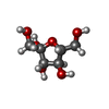  Type: D-saccharide, beta linking / Mass: 180.156 Da / Num. of mol.: 2 / Source method: obtained synthetically / Formula: C6H12O6 / Feature type: SUBJECT OF INVESTIGATION Type: D-saccharide, beta linking / Mass: 180.156 Da / Num. of mol.: 2 / Source method: obtained synthetically / Formula: C6H12O6 / Feature type: SUBJECT OF INVESTIGATION#3: Chemical | ChemComp-CA /   Mass: 40.078 Da / Num. of mol.: 4 / Source method: obtained synthetically / Formula: Ca / Feature type: SUBJECT OF INVESTIGATION Mass: 40.078 Da / Num. of mol.: 4 / Source method: obtained synthetically / Formula: Ca / Feature type: SUBJECT OF INVESTIGATION#4: Water | ChemComp-HOH / |  Mass: 18.015 Da / Num. of mol.: 335 / Source method: isolated from a natural source / Formula: H2O Mass: 18.015 Da / Num. of mol.: 335 / Source method: isolated from a natural source / Formula: H2OHas ligand of interest | Y | Has protein modification | N | |
|---|
-Experimental details
-Experiment
| Experiment | Method: X-RAY DIFFRACTION / Number of used crystals: 1 |
|---|
- Sample preparation
| Crystal | Density Matthews: 1.55 Å3/Da / Density % sol: 20.5 % |
|---|---|
| Crystal grow | Temperature: 289 K / Method: vapor diffusion, sitting drop Details: 0.1 M HEPES pH 7.5 25% w/v Polyethylene glycol 3,350 |
-Data collection
| Diffraction | Mean temperature: 100 K / Serial crystal experiment: N |
|---|---|
| Diffraction source | Source: SYNCHROTRON / Site: SSRF / Beamline: BL19U1 / Wavelength: 0.97854 Å |
| Detector | Type: DECTRIS PILATUS3 6M / Detector: PIXEL / Date: Nov 2, 2023 |
| Radiation | Protocol: SINGLE WAVELENGTH / Monochromatic (M) / Laue (L): M / Scattering type: x-ray |
| Radiation wavelength | Wavelength: 0.97854 Å / Relative weight: 1 |
| Reflection | Resolution: 2.14→50 Å / Num. obs: 59439 / % possible obs: 98.8 % / Redundancy: 11.5 % / CC1/2: 0.997 / Rmerge(I) obs: 0.118 / Rpim(I) all: 0.035 / Rrim(I) all: 0.123 / Χ2: 0.94 / Net I/σ(I): 17 |
| Reflection shell | Resolution: 2.14→2.2 Å / % possible obs: 89.4 % / Redundancy: 6.2 % / Rmerge(I) obs: 0.346 / Num. measured all: 25239 / Num. unique obs: 4085 / CC1/2: 0.944 / Rpim(I) all: 0.143 / Rrim(I) all: 0.375 / Χ2: 0.68 / Net I/σ(I) obs: 5.7 |
- Processing
| Software |
| ||||||||||||||||||||||||||||||||||||||||||||||||||||||||||||||||||||||||||||||||||||||||||||||||||||||||||||||||||||||||||||||||||||||||||||||||||||||||||||||||||||||||||||||||||||||
|---|---|---|---|---|---|---|---|---|---|---|---|---|---|---|---|---|---|---|---|---|---|---|---|---|---|---|---|---|---|---|---|---|---|---|---|---|---|---|---|---|---|---|---|---|---|---|---|---|---|---|---|---|---|---|---|---|---|---|---|---|---|---|---|---|---|---|---|---|---|---|---|---|---|---|---|---|---|---|---|---|---|---|---|---|---|---|---|---|---|---|---|---|---|---|---|---|---|---|---|---|---|---|---|---|---|---|---|---|---|---|---|---|---|---|---|---|---|---|---|---|---|---|---|---|---|---|---|---|---|---|---|---|---|---|---|---|---|---|---|---|---|---|---|---|---|---|---|---|---|---|---|---|---|---|---|---|---|---|---|---|---|---|---|---|---|---|---|---|---|---|---|---|---|---|---|---|---|---|---|---|---|---|---|
| Refinement | Method to determine structure: MOLECULAR REPLACEMENT / Resolution: 2.14→47.54 Å / Cor.coef. Fo:Fc: 0.944 / Cor.coef. Fo:Fc free: 0.912 / SU B: 4.494 / SU ML: 0.119 / Cross valid method: THROUGHOUT / ESU R: 0.239 / ESU R Free: 0.193 / Stereochemistry target values: MAXIMUM LIKELIHOOD / Details: HYDROGENS HAVE BEEN ADDED IN THE RIDING POSITIONS
| ||||||||||||||||||||||||||||||||||||||||||||||||||||||||||||||||||||||||||||||||||||||||||||||||||||||||||||||||||||||||||||||||||||||||||||||||||||||||||||||||||||||||||||||||||||||
| Solvent computation | Ion probe radii: 0.8 Å / Shrinkage radii: 0.8 Å / VDW probe radii: 1.2 Å / Solvent model: MASK | ||||||||||||||||||||||||||||||||||||||||||||||||||||||||||||||||||||||||||||||||||||||||||||||||||||||||||||||||||||||||||||||||||||||||||||||||||||||||||||||||||||||||||||||||||||||
| Displacement parameters | Biso mean: 24.801 Å2
| ||||||||||||||||||||||||||||||||||||||||||||||||||||||||||||||||||||||||||||||||||||||||||||||||||||||||||||||||||||||||||||||||||||||||||||||||||||||||||||||||||||||||||||||||||||||
| Refinement step | Cycle: 1 / Resolution: 2.14→47.54 Å
| ||||||||||||||||||||||||||||||||||||||||||||||||||||||||||||||||||||||||||||||||||||||||||||||||||||||||||||||||||||||||||||||||||||||||||||||||||||||||||||||||||||||||||||||||||||||
| Refine LS restraints |
|