 Movie
Movie Controller


+ Open data

- Basic information
| Entry | Database: PDB / ID: 8xm1 | ||||||
|---|---|---|---|---|---|---|---|
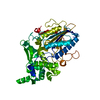
| Title | Phytase mutant APPAmut4 | ||||||
 Components Components | Phytase | ||||||
 Keywords Keywords | HYDROLASE / Phytase mutant APPAmut4 | ||||||
| Function / homology |  Function and homology information Function and homology information4-phytase / inositol hexakisphosphate 4-phosphatase activity / sugar-phosphatase activity / outer membrane-bounded periplasmic space Similarity search - Function | ||||||
| Biological species |  Yersinia intermedia (bacteria) Yersinia intermedia (bacteria) | ||||||
| Method |  X-RAY DIFFRACTION / SYNCHROTRON / MOLECULAR REPLACEMENT / Resolution: 1.9 Å X-RAY DIFFRACTION / SYNCHROTRON / MOLECULAR REPLACEMENT / Resolution: 1.9 Å | ||||||
 Authors Authors | Tu, T. / Wang, Q. | ||||||
| Funding support |  China, 1items China, 1items
| ||||||
 Citation Citation | Journal: J.Biol.Chem. / Year: 2024 Title: Achieving thermostability of a phytase with resistance up to 100 °C. Authors: Tu, T. / Wang, Q. / Dong, R. / Liu, X. / Penttinen, L. / Hakulinen, N. / Tian, J. / Zhang, W. / Wang, Y. / Luo, H. / Yao, B. / Huang, H. #1: Journal: To Be PublishedTitle: Enhancing the thermostability of phytase up to its boiling point through a structured-based computational approach Authors: Tu, T. / Wang, Q. | ||||||
| History |
|
- Structure visualization
| Structure viewer | Molecule: MolmilJmol/JSmol |
|---|
- Downloads & links
-Download
| PDBx/mmCIF format | 8xm1.cif.gz | 226.8 KB | Display | PDBx/mmCIF format |
|---|---|---|---|---|
| PDB format | pdb8xm1.ent.gz | 149 KB | Display | PDB format |
| PDBx/mmJSON format | 8xm1.json.gz | Tree view | PDBx/mmJSON format | |
| Others |  Other downloads Other downloads |
-Validation report
| Arichive directory | https://data.pdbj.org/pub/pdb/validation_reports/xm/8xm1ftp://data.pdbj.org/pub/pdb/validation_reports/xm/8xm1 | HTTPS FTP |
|---|
-Related structure data
-Links
 PDBj
PDBj


- Assembly
| Deposited unit | 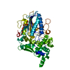
| ||||||||||||
|---|---|---|---|---|---|---|---|---|---|---|---|---|---|
| 1 |
| ||||||||||||
| Unit cell |
|
-Components
| #1: Protein | Mass: 45619.789 Da / Num. of mol.: 1 Source method: isolated from a genetically manipulated source Source: (gene. exp.) Yersinia intermedia (bacteria) / Production host:  Komagataella pastoris (fungus) / References: UniProt: Q000T0, 4-phytase Komagataella pastoris (fungus) / References: UniProt: Q000T0, 4-phytase |
|---|---|
| #2: Chemical | ChemComp-TRS /   Mass: 122.143 Da / Num. of mol.: 1 / Source method: obtained synthetically / Formula: C4H12NO3 / Feature type: SUBJECT OF INVESTIGATION / Comment: pH buffer*YM Mass: 122.143 Da / Num. of mol.: 1 / Source method: obtained synthetically / Formula: C4H12NO3 / Feature type: SUBJECT OF INVESTIGATION / Comment: pH buffer*YM |
| #3: Water | ChemComp-HOH /  Mass: 18.015 Da / Num. of mol.: 401 / Source method: isolated from a natural source / Formula: H2O Mass: 18.015 Da / Num. of mol.: 401 / Source method: isolated from a natural source / Formula: H2O |
| Has ligand of interest | Y |
| Has protein modification | Y |
-Experimental details
-Experiment
| Experiment | Method: X-RAY DIFFRACTION / Number of used crystals: 1 |
|---|
- Sample preparation
| Crystal | Density Matthews: 2.12 Å3/Da / Density % sol: 41.86 % |
|---|---|
| Crystal grow | Temperature: 293 K / Method: vapor diffusion, hanging drop / pH: 4.5 Details: 0.1 M Sodium acetate trihydrate pH 4.5; 30% w/v Polyethylene glycol 1,500 |
-Data collection
| Diffraction | Mean temperature: 100 K / Serial crystal experiment: N |
|---|---|
| Diffraction source | Source: SYNCHROTRON / Site: SSRF / Beamline: BL10U2 / Wavelength: 1 Å |
| Detector | Type: DECTRIS PILATUS 6M / Detector: PIXEL / Date: Jan 14, 2022 |
| Radiation | Protocol: SINGLE WAVELENGTH / Monochromatic (M) / Laue (L): M / Scattering type: x-ray |
| Radiation wavelength | Wavelength: 1 Å / Relative weight: 1 |
| Reflection | Resolution: 1.9→26.35 Å / Num. obs: 31168 / % possible obs: 98.22 % / Redundancy: 2 % / Biso Wilson estimate: 20.12 Å2 / CC1/2: 0.993 / Net I/σ(I): 8.95 |
| Reflection shell | Resolution: 1.9→1.96 Å / Mean I/σ(I) obs: 2.46 / Num. unique obs: 2732 / CC1/2: 0.876 |
- Processing
| Software |
| |||||||||||||||||||||||||||||||||||||||||||||||||||||||||||||||||||||||||||||||||||||||||||
|---|---|---|---|---|---|---|---|---|---|---|---|---|---|---|---|---|---|---|---|---|---|---|---|---|---|---|---|---|---|---|---|---|---|---|---|---|---|---|---|---|---|---|---|---|---|---|---|---|---|---|---|---|---|---|---|---|---|---|---|---|---|---|---|---|---|---|---|---|---|---|---|---|---|---|---|---|---|---|---|---|---|---|---|---|---|---|---|---|---|---|---|---|
| Refinement | Method to determine structure: MOLECULAR REPLACEMENT / Resolution: 1.9→26.35 Å / SU ML: 0.2113 / Cross valid method: FREE R-VALUE / σ(F): 1.33 / Phase error: 20.1005 / Stereochemistry target values: CDL v1.2
| |||||||||||||||||||||||||||||||||||||||||||||||||||||||||||||||||||||||||||||||||||||||||||
| Solvent computation | Shrinkage radii: 0.9 Å / VDW probe radii: 1.11 Å / Solvent model: FLAT BULK SOLVENT MODEL | |||||||||||||||||||||||||||||||||||||||||||||||||||||||||||||||||||||||||||||||||||||||||||
| Displacement parameters | Biso mean: 21.61 Å2 | |||||||||||||||||||||||||||||||||||||||||||||||||||||||||||||||||||||||||||||||||||||||||||
| Refinement step | Cycle: LAST / Resolution: 1.9→26.35 Å
| |||||||||||||||||||||||||||||||||||||||||||||||||||||||||||||||||||||||||||||||||||||||||||
| Refine LS restraints |
| |||||||||||||||||||||||||||||||||||||||||||||||||||||||||||||||||||||||||||||||||||||||||||
| LS refinement shell |
| |||||||||||||||||||||||||||||||||||||||||||||||||||||||||||||||||||||||||||||||||||||||||||
| Refinement TLS params. | Method: refined / Origin x: -6.88853576911 Å / Origin y: 4.4864080037 Å / Origin z: -26.3278598937 Å
| |||||||||||||||||||||||||||||||||||||||||||||||||||||||||||||||||||||||||||||||||||||||||||
| Refinement TLS group | Selection details: all |
