 Movie
Movie Controller


+ Open data

- Basic information
| Entry | Database: PDB / ID: 8x79 | ||||||
|---|---|---|---|---|---|---|---|
| Title | MRE-269 bound Prostacyclin Receptor G protein complex | ||||||
 Components Components |
| ||||||
 Keywords Keywords | SIGNALING PROTEIN / Agonist / Complex / lipid receptor | ||||||
| Function / homology |  Function and homology information Function and homology informationprostacyclin receptor activity / Prostanoid ligand receptors / negative regulation of platelet-derived growth factor receptor signaling pathway / G protein-coupled receptor signaling pathway, coupled to cyclic nucleotide second messenger / guanyl-nucleotide exchange factor activity / negative regulation of smooth muscle cell proliferation / G-protein beta/gamma-subunit complex binding / adenylate cyclase-activating G protein-coupled receptor signaling pathway / Olfactory Signaling Pathway / Activation of the phototransduction cascade ...prostacyclin receptor activity / Prostanoid ligand receptors / negative regulation of platelet-derived growth factor receptor signaling pathway / G protein-coupled receptor signaling pathway, coupled to cyclic nucleotide second messenger / guanyl-nucleotide exchange factor activity / negative regulation of smooth muscle cell proliferation / G-protein beta/gamma-subunit complex binding / adenylate cyclase-activating G protein-coupled receptor signaling pathway / Olfactory Signaling Pathway / Activation of the phototransduction cascade / G beta:gamma signalling through PLC beta / Presynaptic function of Kainate receptors / Thromboxane signalling through TP receptor / G protein-coupled acetylcholine receptor signaling pathway / Activation of G protein gated Potassium channels / Inhibition of voltage gated Ca2+ channels via Gbeta/gamma subunits / G-protein activation / G beta:gamma signalling through CDC42 / Prostacyclin signalling through prostacyclin receptor / Glucagon signaling in metabolic regulation / G beta:gamma signalling through BTK / Synthesis, secretion, and inactivation of Glucagon-like Peptide-1 (GLP-1) / ADP signalling through P2Y purinoceptor 12 / photoreceptor disc membrane / Glucagon-type ligand receptors / Sensory perception of sweet, bitter, and umami (glutamate) taste / Adrenaline,noradrenaline inhibits insulin secretion / Vasopressin regulates renal water homeostasis via Aquaporins / Glucagon-like Peptide-1 (GLP1) regulates insulin secretion / G alpha (z) signalling events / ADP signalling through P2Y purinoceptor 1 / ADORA2B mediated anti-inflammatory cytokines production / cellular response to catecholamine stimulus / G beta:gamma signalling through PI3Kgamma / adenylate cyclase-activating dopamine receptor signaling pathway / Cooperation of PDCL (PhLP1) and TRiC/CCT in G-protein beta folding / cell-cell signaling / GPER1 signaling / G-protein beta-subunit binding / cellular response to prostaglandin E stimulus / heterotrimeric G-protein complex / G alpha (12/13) signalling events / Inactivation, recovery and regulation of the phototransduction cascade / extracellular vesicle / sensory perception of taste / Thrombin signalling through proteinase activated receptors (PARs) / signaling receptor complex adaptor activity / positive regulation of cytosolic calcium ion concentration / retina development in camera-type eye / GTPase binding / Ca2+ pathway / fibroblast proliferation / High laminar flow shear stress activates signaling by PIEZO1 and PECAM1:CDH5:KDR in endothelial cells / response to lipopolysaccharide / G alpha (i) signalling events / G alpha (s) signalling events / phospholipase C-activating G protein-coupled receptor signaling pathway / G alpha (q) signalling events / Ras protein signal transduction / Extra-nuclear estrogen signaling / cell population proliferation / G protein-coupled receptor signaling pathway / inflammatory response / lysosomal membrane / GTPase activity / synapse / GTP binding / protein-containing complex binding / signal transduction / extracellular exosome / metal ion binding / membrane / plasma membrane / cytoplasm / cytosol Similarity search - Function | ||||||
| Biological species |  Homo sapiens (human) Homo sapiens (human) | ||||||
| Method | ELECTRON MICROSCOPY / single particle reconstruction / cryo EM / Resolution: 2.41 Å | ||||||
 Authors Authors | Wang, J.J. / Jin, S. / Zhang, H. / Xu, Y. / Hu, W. / Jiang, Y. / Chen, C. / Wang, D.W. / Xu, H.E. / Wu, C. | ||||||
| Funding support |  China, 1items China, 1items
| ||||||
 Citation Citation | Journal: Sci Adv / Year: 2024 Title: Molecular recognition and activation of the prostacyclin receptor by anti-pulmonary arterial hypertension drugs. Authors: James Jiqi Wang / Sanshan Jin / Heng Zhang / Youwei Xu / Wen Hu / Yi Jiang / Chen Chen / Dao Wen Wang / H Eric Xu / Canrong Wu / Abstract: The prostacyclin (PGI) receptor (IP) is a G-coupled receptor associated with blood pressure regulation, allergy, and inflammatory response. It is a main therapeutic target for pulmonary arterial ...The prostacyclin (PGI) receptor (IP) is a G-coupled receptor associated with blood pressure regulation, allergy, and inflammatory response. It is a main therapeutic target for pulmonary arterial hypertension (PAH) and several other diseases. Here we report cryo-electron microscopy (cryo-EM) structures of the human IP-G complex bound with two anti-PAH drugs, treprostinil and MRE-269 (active form of selexipag), at global resolutions of 2.56 and 2.41 angstrom, respectively. These structures revealed distinct features governing IP ligand binding, receptor activation, and G protein coupling. Moreover, comparison of the activated IP structures uncovered the mechanism and key residues that determine the superior selectivity of MRE-269 over treprostinil. Combined with molecular docking and functional studies, our structures provide insight into agonist selectivity, ligand recognition, receptor activation, and G protein coupling. Our results provide a structural template for further improving IP-targeting drugs to reduce off-target activation of prostanoid receptors and adverse effects. | ||||||
| History |
|
- Structure visualization
| Structure viewer | Molecule: MolmilJmol/JSmol |
|---|
- Downloads & links
-Download
| PDBx/mmCIF format | 8x79.cif.gz | 349.5 KB | Display | PDBx/mmCIF format |
|---|---|---|---|---|
| PDB format | pdb8x79.ent.gz | 283.1 KB | Display | PDB format |
| PDBx/mmJSON format | 8x79.json.gz | Tree view | PDBx/mmJSON format | |
| Others |  Other downloads Other downloads |
-Validation report
| Arichive directory | https://data.pdbj.org/pub/pdb/validation_reports/x7/8x79ftp://data.pdbj.org/pub/pdb/validation_reports/x7/8x79 | HTTPS FTP |
|---|
-Related structure data
| Related structure data |  38095MC  8x7aC M: map data used to model this data C: citing same article ( |
|---|---|
| Similar structure data |
-Links
 PDBj
PDBj


- Assembly
| Deposited unit | 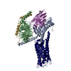
|
|---|---|
| 1 |
|
-Components
-Guanine nucleotide-binding protein ... , 3 types, 3 molecules ABG
| #1: Protein | Mass: 29068.906 Da / Num. of mol.: 1 Source method: isolated from a genetically manipulated source Source: (gene. exp.) Homo sapiens (human) / Gene: GNAS / Production host:   Spodoptera frugiperda (fall armyworm) / References: UniProt: A0A590UJY2 Spodoptera frugiperda (fall armyworm) / References: UniProt: A0A590UJY2 |
|---|---|
| #2: Protein | Mass: 38613.176 Da / Num. of mol.: 1 Source method: isolated from a genetically manipulated source Source: (gene. exp.) Homo sapiens (human) / Gene: GNB1 / Production host: Spodoptera frugiperda (fall armyworm) / References: UniProt: P62873 |
| #3: Protein | Mass: 7861.143 Da / Num. of mol.: 1 Source method: isolated from a genetically manipulated source Source: (gene. exp.) Homo sapiens (human) / Gene: GNG2 / Production host: Spodoptera frugiperda (fall armyworm) / References: UniProt: P59768 |
-Protein , 2 types, 2 molecules NR
| #4: Protein | Mass: 14714.320 Da / Num. of mol.: 1 Source method: isolated from a genetically manipulated source Source: (gene. exp.) Homo sapiens (human) / Production host:  |
|---|---|
| #5: Protein | Mass: 40989.914 Da / Num. of mol.: 1 Source method: isolated from a genetically manipulated source Source: (gene. exp.) Homo sapiens (human) / Gene: PTGIR, PRIPR / Production host: Spodoptera frugiperda (fall armyworm) / References: UniProt: P43119 |
-Non-polymers , 1 types, 1 molecules
| #6: Chemical | ChemComp-Y9Q / Mass: 419.516 Da / Num. of mol.: 1 / Source method: obtained synthetically / Formula: C25H29N3O3 / Feature type: SUBJECT OF INVESTIGATION |
|---|
-Details
| Has ligand of interest | Y |
|---|
-Experimental details
-Experiment
| Experiment | Method: ELECTRON MICROSCOPY |
|---|---|
| EM experiment | Aggregation state: PARTICLE / 3D reconstruction method: single particle reconstruction |
- Sample preparation
| Component | Name: IP-Gs complex / Type: COMPLEX / Entity ID: #5, #1-#4 / Source: RECOMBINANT |
|---|---|
| Molecular weight | Units: KILODALTONS/NANOMETER / Experimental value: NO |
| Source (natural) | Organism: Homo sapiens (human) |
| Source (recombinant) | Organism: Spodoptera frugiperda (fall armyworm) |
| Buffer solution | pH: 7.5 |
| Specimen | Embedding applied: NO / Shadowing applied: NO / Staining applied: NO / Vitrification applied: YES |
| Vitrification | Cryogen name: ETHANE |
- Electron microscopy imaging
| Experimental equipment |  Model: Titan Krios / Image courtesy: FEI Company |
|---|---|
| Microscopy | Model: FEI TITAN KRIOS |
| Electron gun | Electron source:  FIELD EMISSION GUN / Accelerating voltage: 300 kV / Illumination mode: OTHER FIELD EMISSION GUN / Accelerating voltage: 300 kV / Illumination mode: OTHER |
| Electron lens | Mode: BRIGHT FIELD / Nominal defocus max: 5000 nm / Nominal defocus min: 1200 nm |
| Image recording | Electron dose: 50 e/Å2 / Film or detector model: FEI FALCON IV (4k x 4k) |
- Processing
| CTF correction | Type: NONE |
|---|---|
| 3D reconstruction | Resolution: 2.41 Å / Resolution method: FSC 0.143 CUT-OFF / Num. of particles: 206075 / Symmetry type: POINT |
| Atomic model building | Protocol: OTHER |
