+ Open data
- Basic information
| Entry | Database: PDB / ID: 8x77 | ||||||
|---|---|---|---|---|---|---|---|
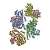
| Title | Enterovirus proteinase with host factor | ||||||
 Components Components |
| ||||||
 Keywords Keywords | CELL INVASION / host protein / VIRAL PROTEIN | ||||||
| Function / homology |  Function and homology information Function and homology informationpeptidyl-histidine methylation / regulation of uterine smooth muscle contraction / protein-histidine N-methyltransferase / protein-L-histidine N-tele-methyltransferase activity / actin modification / histone H3K36 methyltransferase activity / histone H3K4 methyltransferase activity / positive regulation of muscle cell differentiation / viral process / cysteine-type peptidase activity ...peptidyl-histidine methylation / regulation of uterine smooth muscle contraction / protein-histidine N-methyltransferase / protein-L-histidine N-tele-methyltransferase activity / actin modification / histone H3K36 methyltransferase activity / histone H3K4 methyltransferase activity / positive regulation of muscle cell differentiation / viral process / cysteine-type peptidase activity / PKMTs methylate histone lysines / virion component / actin binding / RNA polymerase II-specific DNA-binding transcription factor binding / host cell cytoplasm / transcription coactivator activity / chromatin / positive regulation of DNA-templated transcription / positive regulation of transcription by RNA polymerase II / proteolysis / nucleoplasm / metal ion binding / cytoplasm Similarity search - Function | ||||||
| Biological species |  Homo sapiens (human) Homo sapiens (human)  Enterovirus A71 Enterovirus A71 | ||||||
| Method |  X-RAY DIFFRACTION / X-RAY DIFFRACTION /  SYNCHROTRON / SYNCHROTRON /  MOLECULAR REPLACEMENT / Resolution: 3.52 Å MOLECULAR REPLACEMENT / Resolution: 3.52 Å | ||||||
 Authors Authors | Gao, X. / Cui, S. | ||||||
| Funding support | 1items
| ||||||
 Citation Citation |  Journal: Nat Commun / Year: 2024 Journal: Nat Commun / Year: 2024Title: The EV71 2A protease occupies the central cleft of SETD3 and disrupts SETD3-actin interaction. Authors: Xiaopan Gao / Bei Wang / Kaixiang Zhu / Linyue Wang / Bo Qin / Kun Shang / Wei Ding / Jianwei Wang / Sheng Cui /  Abstract: SETD3 is an essential host factor for the replication of a variety of enteroviruses that specifically interacts with viral protease 2A. However, the interaction between SETD3 and the 2A protease has ...SETD3 is an essential host factor for the replication of a variety of enteroviruses that specifically interacts with viral protease 2A. However, the interaction between SETD3 and the 2A protease has not been fully characterized. Here, we use X-ray crystallography and cryo-electron microscopy to determine the structures of SETD3 complexed with the 2A protease of EV71 to 3.5 Å and 3.1 Å resolution, respectively. We find that the 2A protease occupies the V-shaped central cleft of SETD3 through two discrete sites. The relative positions of the two proteins vary in the crystal and cryo-EM structures, showing dynamic binding. A biolayer interferometry assay shows that the EV71 2A protease outcompetes actin for SETD3 binding. We identify key 2A residues involved in SETD3 binding and demonstrate that 2A's ability to bind SETD3 correlates with EV71 production in cells. Coimmunoprecipitation experiments in EV71 infected and 2A expressing cells indicate that 2A interferes with the SETD3-actin complex, and the disruption of this complex reduces enterovirus replication. Together, these results reveal the molecular mechanism underlying the interplay between SETD3, actin, and viral 2A during virus replication. | ||||||
| History |
|
- Structure visualization
| Structure viewer | Molecule:  Molmil Molmil Jmol/JSmol Jmol/JSmol |
|---|
- Downloads & links
- Download
| PDBx/mmCIF format |  8x77.cif.gz 8x77.cif.gz | 545.1 KB | Display |  PDBx/mmCIF format PDBx/mmCIF format |
|---|---|---|---|---|
| PDB format |  pdb8x77.ent.gz pdb8x77.ent.gz | 434.1 KB | Display |  PDB format PDB format |
| PDBx/mmJSON format |  8x77.json.gz 8x77.json.gz | Tree view |  PDBx/mmJSON format PDBx/mmJSON format | |
| Others |  Other downloads Other downloads |
-Validation report
| Summary document |  8x77_validation.pdf.gz 8x77_validation.pdf.gz | 1.6 MB | Display |  wwPDB validaton report wwPDB validaton report |
|---|---|---|---|---|
| Full document |  8x77_full_validation.pdf.gz 8x77_full_validation.pdf.gz | 1.7 MB | Display | |
| Data in XML |  8x77_validation.xml.gz 8x77_validation.xml.gz | 111.8 KB | Display | |
| Data in CIF |  8x77_validation.cif.gz 8x77_validation.cif.gz | 157.5 KB | Display | |
| Arichive directory |  https://data.pdbj.org/pub/pdb/validation_reports/x7/8x77 https://data.pdbj.org/pub/pdb/validation_reports/x7/8x77 ftp://data.pdbj.org/pub/pdb/validation_reports/x7/8x77 ftp://data.pdbj.org/pub/pdb/validation_reports/x7/8x77 | HTTPS FTP |
-Related structure data
| Related structure data |  8x8qC C: citing same article ( |
|---|---|
| Similar structure data | Similarity search - Function & homology  F&H Search F&H Search |
- Links
Links
- Assembly
| Deposited unit | 
| ||||||||
|---|---|---|---|---|---|---|---|---|---|
| 1 | 
| ||||||||
| 2 | 
| ||||||||
| 3 | 
| ||||||||
| 4 | 
| ||||||||
| Unit cell |
|
- Components
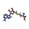
| #1: Protein | Mass: 67342.047 Da / Num. of mol.: 4 Source method: isolated from a genetically manipulated source Source: (gene. exp.)  Homo sapiens (human) / Gene: SETD3 / Production host: Homo sapiens (human) / Gene: SETD3 / Production host:  Baculovirus expression vector pFastBac1-HM / References: UniProt: Q86TU7 Baculovirus expression vector pFastBac1-HM / References: UniProt: Q86TU7#2: Protein | Mass: 16562.418 Da / Num. of mol.: 4 / Mutation: C110A Source method: isolated from a genetically manipulated source Source: (gene. exp.)   Enterovirus A71 / Production host: Enterovirus A71 / Production host:  Baculovirus expression vector pFastBac1-HM / References: UniProt: R9YK28 Baculovirus expression vector pFastBac1-HM / References: UniProt: R9YK28#3: Chemical | ChemComp-SAH / | #4: Chemical | ChemComp-ZN / #5: Water | ChemComp-HOH / | Has ligand of interest | Y | |
|---|
-Experimental details
-Experiment
| Experiment | Method:  X-RAY DIFFRACTION / Number of used crystals: 1 X-RAY DIFFRACTION / Number of used crystals: 1 |
|---|
- Sample preparation
| Crystal | Density Matthews: 2.37 Å3/Da / Density % sol: 48.04 % |
|---|---|
| Crystal grow | Temperature: 296 K / Method: vapor diffusion, hanging drop Details: 0.02M Citric acid, 0.08M BIS-TRIS propane, pH8.8, 16% PEG 3350 |
-Data collection
| Diffraction | Mean temperature: 100 K / Serial crystal experiment: N |
|---|---|
| Diffraction source | Source:  SYNCHROTRON / Site: SYNCHROTRON / Site:  SSRF SSRF  / Beamline: BL17B1 / Wavelength: 0.9785 Å / Beamline: BL17B1 / Wavelength: 0.9785 Å |
| Detector | Type: DECTRIS PILATUS3 S 6M / Detector: PIXEL / Date: Jul 3, 2020 |
| Radiation | Protocol: SINGLE WAVELENGTH / Monochromatic (M) / Laue (L): M / Scattering type: x-ray |
| Radiation wavelength | Wavelength: 0.9785 Å / Relative weight: 1 |
| Reflection twin | Operator: h,-k,-h-l / Fraction: 0.09 |
| Reflection | Resolution: 3.5→49.34 Å / Num. obs: 76091 / % possible obs: 97.9 % / Redundancy: 3.51 % / CC1/2: 0.93 / Net I/σ(I): 2.29 |
| Reflection shell | Resolution: 3.5→3.51 Å / Rmerge(I) obs: 2.09 / Num. unique obs: 11533 |
- Processing
| Software |
| |||||||||||||||||||||||||||||||||||||||||||||||||||||||||||||||||||||||||||||||||||||||||||||||||||||||||
|---|---|---|---|---|---|---|---|---|---|---|---|---|---|---|---|---|---|---|---|---|---|---|---|---|---|---|---|---|---|---|---|---|---|---|---|---|---|---|---|---|---|---|---|---|---|---|---|---|---|---|---|---|---|---|---|---|---|---|---|---|---|---|---|---|---|---|---|---|---|---|---|---|---|---|---|---|---|---|---|---|---|---|---|---|---|---|---|---|---|---|---|---|---|---|---|---|---|---|---|---|---|---|---|---|---|---|
| Refinement | Method to determine structure:  MOLECULAR REPLACEMENT / Resolution: 3.52→19.99 Å / Cross valid method: THROUGHOUT / σ(F): 0 / Phase error: 31.23 / Stereochemistry target values: TWIN_LSQ_F MOLECULAR REPLACEMENT / Resolution: 3.52→19.99 Å / Cross valid method: THROUGHOUT / σ(F): 0 / Phase error: 31.23 / Stereochemistry target values: TWIN_LSQ_FDetails: There are 4 pairs of molecules in the ASU. Only chains A/C have been conducted in-depth analysis and optimization and used for further structure analysis. Other molecules have poor electron ...Details: There are 4 pairs of molecules in the ASU. Only chains A/C have been conducted in-depth analysis and optimization and used for further structure analysis. Other molecules have poor electron density and were not carried out in-depth optimization.
| |||||||||||||||||||||||||||||||||||||||||||||||||||||||||||||||||||||||||||||||||||||||||||||||||||||||||
| Solvent computation | Shrinkage radii: 0.9 Å / VDW probe radii: 1.11 Å / Solvent model: FLAT BULK SOLVENT MODEL | |||||||||||||||||||||||||||||||||||||||||||||||||||||||||||||||||||||||||||||||||||||||||||||||||||||||||
| Displacement parameters | Biso max: 231.48 Å2 / Biso mean: 90.9712 Å2 / Biso min: 0.23 Å2 | |||||||||||||||||||||||||||||||||||||||||||||||||||||||||||||||||||||||||||||||||||||||||||||||||||||||||
| Refinement step | Cycle: final / Resolution: 3.52→19.99 Å
| |||||||||||||||||||||||||||||||||||||||||||||||||||||||||||||||||||||||||||||||||||||||||||||||||||||||||
| LS refinement shell | Refine-ID: X-RAY DIFFRACTION / Rfactor Rfree error: 0 / Total num. of bins used: 14
|
 Movie
Movie Controller


 PDBj
PDBj


