 Movie
Movie Controller

[English] 日本語
 Yorodumi
- PDB-8x6m: Crystal Structure of Glycerol Dehydrogenase in the Presence of NA... -
+ Open data

- Basic information
| Entry | Database: PDB / ID: 8x6m | ||||||
|---|---|---|---|---|---|---|---|
| Title | Crystal Structure of Glycerol Dehydrogenase in the Presence of NAD+ and Glycerol | ||||||
 Components Components | Glycerol dehydrogenase | ||||||
 Keywords Keywords | OXIDOREDUCTASE / Glycerol Dehydrogenase / Complex | ||||||
| Function / homology |  Function and homology information Function and homology information(R)-aminopropanol dehydrogenase / (R)-aminopropanol dehydrogenase activity / anaerobic glycerol catabolic process / methylglyoxal catabolic process / glycerol dehydrogenase / glycerol dehydrogenase (NAD+) activity / protein homotetramerization / protein-containing complex / zinc ion binding / identical protein binding / cytosol Similarity search - Function | ||||||
| Biological species |  | ||||||
| Method |  X-RAY DIFFRACTION / SYNCHROTRON / MOLECULAR REPLACEMENT / Resolution: 2 Å X-RAY DIFFRACTION / SYNCHROTRON / MOLECULAR REPLACEMENT / Resolution: 2 Å | ||||||
 Authors Authors | Park, T. / Kang, J.Y. / Jin, M. / Yang, J. / Kim, H. / Noh, C. / Eom, S.H. | ||||||
| Funding support |  Korea, Republic Of, 1items Korea, Republic Of, 1items
| ||||||
 Citation Citation | Journal: Plos One / Year: 2024 Title: Structural insights into the octamerization of glycerol dehydrogenase. Authors: Park, T. / Kang, J.Y. / Jin, M. / Yang, J. / Kim, H. / Noh, C. / Jung, C.H. / Eom, S.H. | ||||||
| History |
|
- Structure visualization
| Structure viewer | Molecule: MolmilJmol/JSmol |
|---|
- Downloads & links
-Download
| PDBx/mmCIF format | 8x6m.cif.gz | 158.4 KB | Display | PDBx/mmCIF format |
|---|---|---|---|---|
| PDB format | pdb8x6m.ent.gz | 123.8 KB | Display | PDB format |
| PDBx/mmJSON format | 8x6m.json.gz | Tree view | PDBx/mmJSON format | |
| Others |  Other downloads Other downloads |
-Validation report
| Arichive directory | https://data.pdbj.org/pub/pdb/validation_reports/x6/8x6mftp://data.pdbj.org/pub/pdb/validation_reports/x6/8x6m | HTTPS FTP |
|---|
-Related structure data
| Related structure data | 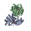 8gobS S: Starting model for refinement |
|---|---|
| Similar structure data |
-Links
 PDBj
PDBj- Assembly
| Deposited unit | 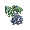
| ||||||||
|---|---|---|---|---|---|---|---|---|---|
| 1 | 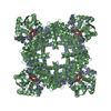
| ||||||||
| Unit cell |
|
-Components
| #1: Protein | Mass: 39817.152 Da / Num. of mol.: 2 Source method: isolated from a genetically manipulated source Source: (gene. exp.) References: UniProt: P0A9S5, glycerol dehydrogenase, (R)-aminopropanol dehydrogenase #2: Chemical |   Mass: 65.409 Da / Num. of mol.: 2 / Source method: isolated from a natural source / Formula: Zn / Feature type: SUBJECT OF INVESTIGATION Mass: 65.409 Da / Num. of mol.: 2 / Source method: isolated from a natural source / Formula: Zn / Feature type: SUBJECT OF INVESTIGATION#3: Chemical |   Mass: 663.425 Da / Num. of mol.: 2 / Source method: obtained synthetically / Formula: C21H27N7O14P2 / Feature type: SUBJECT OF INVESTIGATION / Comment: NAD*YM Mass: 663.425 Da / Num. of mol.: 2 / Source method: obtained synthetically / Formula: C21H27N7O14P2 / Feature type: SUBJECT OF INVESTIGATION / Comment: NAD*YM#4: Chemical |   Mass: 92.094 Da / Num. of mol.: 2 / Source method: obtained synthetically / Formula: C3H8O3 / Feature type: SUBJECT OF INVESTIGATION Mass: 92.094 Da / Num. of mol.: 2 / Source method: obtained synthetically / Formula: C3H8O3 / Feature type: SUBJECT OF INVESTIGATION#5: Water | ChemComp-HOH / |  Mass: 18.015 Da / Num. of mol.: 429 / Source method: isolated from a natural source / Formula: H2O Mass: 18.015 Da / Num. of mol.: 429 / Source method: isolated from a natural source / Formula: H2OHas ligand of interest | Y | |
|---|
-Experimental details
-Experiment
| Experiment | Method: X-RAY DIFFRACTION / Number of used crystals: 1 |
|---|
- Sample preparation
| Crystal | Density Matthews: 3.62 Å3/Da / Density % sol: 66.04 % |
|---|---|
| Crystal grow | Temperature: 293 K / Method: vapor diffusion, hanging drop Details: 100 mM Na2HPO4-citrate (pH 4.2), 200 mM NaCl, and 10% (w/v) PEG 3000 |
-Data collection
| Diffraction | Mean temperature: 100 K / Serial crystal experiment: N |
|---|---|
| Diffraction source | Source: SYNCHROTRON / Site: PAL/PLS / Beamline: 11C / Wavelength: 0.97942 Å |
| Detector | Type: DECTRIS PILATUS3 6M / Detector: PIXEL / Date: Dec 22, 2022 |
| Radiation | Protocol: SINGLE WAVELENGTH / Monochromatic (M) / Laue (L): M / Scattering type: x-ray |
| Radiation wavelength | Wavelength: 0.97942 Å / Relative weight: 1 |
| Reflection | Resolution: 2→118.1 Å / Num. obs: 78784 / % possible obs: 99.9 % / Redundancy: 26.1 % / CC1/2: 0.999 / Net I/σ(I): 19 |
| Reflection shell | Resolution: 2→2 Å / Num. unique obs: 3865 / CC1/2: 0.94 / % possible all: 99.4 |
- Processing
| Software |
| ||||||||||||||||||||||||||||||||||||||||||||||||||||||||||||||||||||||||||||||||||||||||||||||||||||||||||||||||||||||||||||||||||||||||||||||||||||||||||||||||||||||||||||||||||||||
|---|---|---|---|---|---|---|---|---|---|---|---|---|---|---|---|---|---|---|---|---|---|---|---|---|---|---|---|---|---|---|---|---|---|---|---|---|---|---|---|---|---|---|---|---|---|---|---|---|---|---|---|---|---|---|---|---|---|---|---|---|---|---|---|---|---|---|---|---|---|---|---|---|---|---|---|---|---|---|---|---|---|---|---|---|---|---|---|---|---|---|---|---|---|---|---|---|---|---|---|---|---|---|---|---|---|---|---|---|---|---|---|---|---|---|---|---|---|---|---|---|---|---|---|---|---|---|---|---|---|---|---|---|---|---|---|---|---|---|---|---|---|---|---|---|---|---|---|---|---|---|---|---|---|---|---|---|---|---|---|---|---|---|---|---|---|---|---|---|---|---|---|---|---|---|---|---|---|---|---|---|---|---|---|
| Refinement | Method to determine structure: MOLECULAR REPLACEMENT Starting model: 8GOB Resolution: 2→50.01 Å / Cor.coef. Fo:Fc: 0.966 / Cor.coef. Fo:Fc free: 0.945 / SU B: 3.869 / SU ML: 0.1 / Cross valid method: THROUGHOUT / ESU R: 0.123 / ESU R Free: 0.121 / Stereochemistry target values: MAXIMUM LIKELIHOOD / Details: HYDROGENS HAVE BEEN ADDED IN THE RIDING POSITIONS
| ||||||||||||||||||||||||||||||||||||||||||||||||||||||||||||||||||||||||||||||||||||||||||||||||||||||||||||||||||||||||||||||||||||||||||||||||||||||||||||||||||||||||||||||||||||||
| Solvent computation | Ion probe radii: 0.8 Å / Shrinkage radii: 0.8 Å / VDW probe radii: 1.2 Å / Solvent model: MASK | ||||||||||||||||||||||||||||||||||||||||||||||||||||||||||||||||||||||||||||||||||||||||||||||||||||||||||||||||||||||||||||||||||||||||||||||||||||||||||||||||||||||||||||||||||||||
| Displacement parameters | Biso mean: 35.575 Å2
| ||||||||||||||||||||||||||||||||||||||||||||||||||||||||||||||||||||||||||||||||||||||||||||||||||||||||||||||||||||||||||||||||||||||||||||||||||||||||||||||||||||||||||||||||||||||
| Refinement step | Cycle: 1 / Resolution: 2→50.01 Å
| ||||||||||||||||||||||||||||||||||||||||||||||||||||||||||||||||||||||||||||||||||||||||||||||||||||||||||||||||||||||||||||||||||||||||||||||||||||||||||||||||||||||||||||||||||||||
| Refine LS restraints |
|