 Movie
Movie Controller


+ Open data

- Basic information
| Entry | Database: PDB / ID: 8x4q | ||||||
|---|---|---|---|---|---|---|---|
| Title | Apo structure of L-tryptophan specific decarboxylase PsiD | ||||||
 Components Components | (L-tryptophan decarboxylase) x 2 | ||||||
 Keywords Keywords | OXIDOREDUCTASE / decarboxylase / psilocybin / L-tryptophan / psilocin / tryptamine | ||||||
| Function / homology |  Function and homology information Function and homology informationL-tryptophan decarboxylase / L-tryptophan decarboxylase activity / psilocybin biosynthetic process / phosphatidylserine decarboxylase activity / phosphatidylethanolamine biosynthetic process / mitochondrion Similarity search - Function | ||||||
| Biological species |  Psilocybe cubensis (magic mushroom) Psilocybe cubensis (magic mushroom) | ||||||
| Method |  X-RAY DIFFRACTION / SYNCHROTRON / MOLECULAR REPLACEMENT / Resolution: 2.55 Å X-RAY DIFFRACTION / SYNCHROTRON / MOLECULAR REPLACEMENT / Resolution: 2.55 Å | ||||||
 Authors Authors | Meng, C.Y. / Wen, Y. / Guo, W.T. / Wu, B.X. | ||||||
| Funding support | 1items
| ||||||
 Citation Citation | Journal: Nat Commun / Year: 2025 Title: Structural basis for psilocybin biosynthesis. Authors: Meng, C. / Guo, W. / Xiao, C. / Wen, Y. / Zhu, X. / Zhang, Q. / Liang, Y. / Li, H. / Xu, S. / Qiu, Y. / Chen, H. / Lin, W.J. / Wu, B. | ||||||
| History |
|
- Structure visualization
| Structure viewer | Molecule: MolmilJmol/JSmol |
|---|
- Downloads & links
-Download
| PDBx/mmCIF format | 8x4q.cif.gz | 1.1 MB | Display | PDBx/mmCIF format |
|---|---|---|---|---|
| PDB format | pdb8x4q.ent.gz | 751.7 KB | Display | PDB format |
| PDBx/mmJSON format | 8x4q.json.gz | Tree view | PDBx/mmJSON format | |
| Others |  Other downloads Other downloads |
-Validation report
| Arichive directory | https://data.pdbj.org/pub/pdb/validation_reports/x4/8x4qftp://data.pdbj.org/pub/pdb/validation_reports/x4/8x4q | HTTPS FTP |
|---|
-Related structure data
| Related structure data |  8x4sC 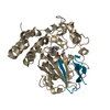 8ziaC  8zicC  8zieC  8zigC  8zihC  8ziiC  8zimC  8zioC C: citing same article ( |
|---|---|
| Similar structure data |
-Links
 PDBj
PDBj- Assembly
| Deposited unit | 
| ||||||||||||
|---|---|---|---|---|---|---|---|---|---|---|---|---|---|
| 1 | 
| ||||||||||||
| 2 | 
| ||||||||||||
| 3 | 
| ||||||||||||
| 4 | 
| ||||||||||||
| 5 | 
| ||||||||||||
| 6 | 
| ||||||||||||
| Unit cell |
|
-Components
| #1: Protein | Mass: 45684.344 Da / Num. of mol.: 6 Source method: isolated from a genetically manipulated source Source: (gene. exp.) Psilocybe cubensis (magic mushroom) / Gene: psiD / Production host:  #2: Protein/peptide | Mass: 4021.680 Da / Num. of mol.: 6 Source method: isolated from a genetically manipulated source Details: The S403 was processed to a pyruvoyl group / Source: (gene. exp.) Psilocybe cubensis (magic mushroom) / Gene: psiD / Production host: #3: Water | ChemComp-HOH / |  Mass: 18.015 Da / Num. of mol.: 868 / Source method: isolated from a natural source / Formula: H2O Mass: 18.015 Da / Num. of mol.: 868 / Source method: isolated from a natural source / Formula: H2OHas ligand of interest | Y | Has protein modification | Y | |
|---|
-Experimental details
-Experiment
| Experiment | Method: X-RAY DIFFRACTION / Number of used crystals: 1 |
|---|
- Sample preparation
| Crystal | Density Matthews: 2.11 Å3/Da / Density % sol: 41.72 % |
|---|---|
| Crystal grow | Temperature: 293 K / Method: vapor diffusion, sitting drop Details: 0.2 M Ammonium acetate, 0.1 M Sodium citrate pH 5.6, 30% w/v Polyethylene glycol 4,000 |
-Data collection
| Diffraction | Mean temperature: 100 K / Serial crystal experiment: N |
|---|---|
| Diffraction source | Source: SYNCHROTRON / Site: SSRF  / Beamline: BL18U1 / Wavelength: 0.97853 Å / Beamline: BL18U1 / Wavelength: 0.97853 Å |
| Detector | Type: DECTRIS PILATUS 6M / Detector: PIXEL / Date: Jul 10, 2023 |
| Radiation | Protocol: SINGLE WAVELENGTH / Monochromatic (M) / Laue (L): M / Scattering type: x-ray |
| Radiation wavelength | Wavelength: 0.97853 Å / Relative weight: 1 |
| Reflection | Resolution: 2.55→30 Å / Num. obs: 80722 / % possible obs: 99.9 % / Redundancy: 6.6 % / Biso Wilson estimate: 37.49 Å2 / CC1/2: 0.992 / Rmerge(I) obs: 0.168 / Rpim(I) all: 0.07 / Rrim(I) all: 0.182 / Net I/σ(I): 9.1 |
| Reflection shell | Resolution: 2.55→2.69 Å / Redundancy: 6.6 % / Rmerge(I) obs: 0.808 / Mean I/σ(I) obs: 2.2 / Num. unique obs: 11754 / CC1/2: 0.755 / Rpim(I) all: 0.34 / Rrim(I) all: 0.878 / % possible all: 100 |
- Processing
| Software |
| ||||||||||||||||||||||||||||||||||||||||||||||||||||||||||||||||||||||||||||||||||||||||||||||||||||||||||||||||||||||||||||||||||||||||||||||||||||||||||||||||||||||||||||||||||||||||||||||||||||||||||||||||||
|---|---|---|---|---|---|---|---|---|---|---|---|---|---|---|---|---|---|---|---|---|---|---|---|---|---|---|---|---|---|---|---|---|---|---|---|---|---|---|---|---|---|---|---|---|---|---|---|---|---|---|---|---|---|---|---|---|---|---|---|---|---|---|---|---|---|---|---|---|---|---|---|---|---|---|---|---|---|---|---|---|---|---|---|---|---|---|---|---|---|---|---|---|---|---|---|---|---|---|---|---|---|---|---|---|---|---|---|---|---|---|---|---|---|---|---|---|---|---|---|---|---|---|---|---|---|---|---|---|---|---|---|---|---|---|---|---|---|---|---|---|---|---|---|---|---|---|---|---|---|---|---|---|---|---|---|---|---|---|---|---|---|---|---|---|---|---|---|---|---|---|---|---|---|---|---|---|---|---|---|---|---|---|---|---|---|---|---|---|---|---|---|---|---|---|---|---|---|---|---|---|---|---|---|---|---|---|---|---|---|---|---|
| Refinement | Method to determine structure: MOLECULAR REPLACEMENT Starting model: AlphaFold Resolution: 2.55→29.37 Å / SU ML: 0.3198 / Cross valid method: FREE R-VALUE / σ(F): 1.34 / Phase error: 23.0393 Stereochemistry target values: GeoStd + Monomer Library + CDL v1.2
| ||||||||||||||||||||||||||||||||||||||||||||||||||||||||||||||||||||||||||||||||||||||||||||||||||||||||||||||||||||||||||||||||||||||||||||||||||||||||||||||||||||||||||||||||||||||||||||||||||||||||||||||||||
| Solvent computation | Shrinkage radii: 0.9 Å / VDW probe radii: 1.1 Å / Solvent model: FLAT BULK SOLVENT MODEL | ||||||||||||||||||||||||||||||||||||||||||||||||||||||||||||||||||||||||||||||||||||||||||||||||||||||||||||||||||||||||||||||||||||||||||||||||||||||||||||||||||||||||||||||||||||||||||||||||||||||||||||||||||
| Displacement parameters | Biso mean: 34.41 Å2 | ||||||||||||||||||||||||||||||||||||||||||||||||||||||||||||||||||||||||||||||||||||||||||||||||||||||||||||||||||||||||||||||||||||||||||||||||||||||||||||||||||||||||||||||||||||||||||||||||||||||||||||||||||
| Refinement step | Cycle: LAST / Resolution: 2.55→29.37 Å
| ||||||||||||||||||||||||||||||||||||||||||||||||||||||||||||||||||||||||||||||||||||||||||||||||||||||||||||||||||||||||||||||||||||||||||||||||||||||||||||||||||||||||||||||||||||||||||||||||||||||||||||||||||
| Refine LS restraints |
| ||||||||||||||||||||||||||||||||||||||||||||||||||||||||||||||||||||||||||||||||||||||||||||||||||||||||||||||||||||||||||||||||||||||||||||||||||||||||||||||||||||||||||||||||||||||||||||||||||||||||||||||||||
| LS refinement shell |
| ||||||||||||||||||||||||||||||||||||||||||||||||||||||||||||||||||||||||||||||||||||||||||||||||||||||||||||||||||||||||||||||||||||||||||||||||||||||||||||||||||||||||||||||||||||||||||||||||||||||||||||||||||
| Refinement TLS params. | Method: refined / Origin x: -22.7452443195 Å / Origin y: -3.02823465971 Å / Origin z: 32.9501162156 Å
| ||||||||||||||||||||||||||||||||||||||||||||||||||||||||||||||||||||||||||||||||||||||||||||||||||||||||||||||||||||||||||||||||||||||||||||||||||||||||||||||||||||||||||||||||||||||||||||||||||||||||||||||||||
| Refinement TLS group | Selection details: all |
