 Movie
Movie Controller


+ Open data

- Basic information
| Entry | Database: PDB / ID: 8wvb | ||||||
|---|---|---|---|---|---|---|---|
| Title | Crystal structure of Lsd18 mutant S195M | ||||||
 Components Components | Putative epoxidase LasC | ||||||
 Keywords Keywords | FLAVOPROTEIN / FAD dependent monooxygenase / epoxidase / mutant | ||||||
| Function / homology | Oxidoreductases; Acting on paired donors, with incorporation or reduction of molecular oxygen; With NADH or NADPH as one donor, and incorporation of one atom of oxygen into the other donor / FAD-binding domain / FAD binding domain / antibiotic biosynthetic process / FAD binding / monooxygenase activity / FAD/NAD(P)-binding domain superfamily / FLAVIN-ADENINE DINUCLEOTIDE / Putative epoxidase LasC Function and homology information Function and homology information | ||||||
| Biological species |  Streptomyces lasalocidi (bacteria) Streptomyces lasalocidi (bacteria) | ||||||
| Method |  X-RAY DIFFRACTION / SYNCHROTRON / MOLECULAR REPLACEMENT / Resolution: 2.5 Å X-RAY DIFFRACTION / SYNCHROTRON / MOLECULAR REPLACEMENT / Resolution: 2.5 Å | ||||||
 Authors Authors | Liu, N. / Xiao, H.L. / Chen, X. | ||||||
| Funding support |  China, 1items China, 1items
| ||||||
 Citation Citation | Journal: Int J Mol Sci / Year: 2023 Title: Simultaneous Improvement in the Thermostability and Catalytic Activity of Epoxidase Lsd18 for the Synthesis of Lasalocid A. Authors: Liu, N. / Xiao, H. / Zang, Y. / Zhou, L. / Mencius, J. / Yang, Z. / Quan, S. / Chen, X. | ||||||
| History |
|
- Structure visualization
| Structure viewer | Molecule: MolmilJmol/JSmol |
|---|
- Downloads & links
-Download
| PDBx/mmCIF format | 8wvb.cif.gz | 348.4 KB | Display | PDBx/mmCIF format |
|---|---|---|---|---|
| PDB format | pdb8wvb.ent.gz | 280.2 KB | Display | PDB format |
| PDBx/mmJSON format | 8wvb.json.gz | Tree view | PDBx/mmJSON format | |
| Others |  Other downloads Other downloads |
-Validation report
| Summary document | 8wvb_validation.pdf.gz | 975.5 KB | Display | wwPDB validaton report |
|---|---|---|---|---|
| Full document | 8wvb_full_validation.pdf.gz | 982.6 KB | Display | |
| Data in XML | 8wvb_validation.xml.gz | 34.4 KB | Display | |
| Data in CIF | 8wvb_validation.cif.gz | 47.8 KB | Display | |
| Arichive directory | https://data.pdbj.org/pub/pdb/validation_reports/wv/8wvbftp://data.pdbj.org/pub/pdb/validation_reports/wv/8wvb | HTTPS FTP |
-Related structure data
| Related structure data | 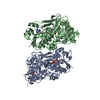 8wvfC  8up4S S: Starting model for refinement C: citing same article ( |
|---|---|
| Similar structure data |
-Links
 PDBj
PDBj- Assembly
| Deposited unit | 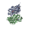
| ||||||||
|---|---|---|---|---|---|---|---|---|---|
| 1 | 
| ||||||||
| 2 | 
| ||||||||
| Unit cell |
|
-Components
| #1: Protein | Mass: 52690.812 Da / Num. of mol.: 2 / Mutation: S195M Source method: isolated from a genetically manipulated source Source: (gene. exp.) Streptomyces lasalocidi (bacteria) / Gene: lsd18 / Production host: #2: Chemical |   Mass: 785.550 Da / Num. of mol.: 2 / Source method: obtained synthetically / Formula: C27H33N9O15P2 / Feature type: SUBJECT OF INVESTIGATION / Comment: FAD*YM Mass: 785.550 Da / Num. of mol.: 2 / Source method: obtained synthetically / Formula: C27H33N9O15P2 / Feature type: SUBJECT OF INVESTIGATION / Comment: FAD*YM#3: Chemical |   Mass: 35.453 Da / Num. of mol.: 2 / Source method: obtained synthetically / Formula: Cl Mass: 35.453 Da / Num. of mol.: 2 / Source method: obtained synthetically / Formula: Cl#4: Water | ChemComp-HOH / |  Mass: 18.015 Da / Num. of mol.: 185 / Source method: isolated from a natural source / Formula: H2O Mass: 18.015 Da / Num. of mol.: 185 / Source method: isolated from a natural source / Formula: H2OHas ligand of interest | Y | |
|---|
-Experimental details
-Experiment
| Experiment | Method: X-RAY DIFFRACTION / Number of used crystals: 1 |
|---|
- Sample preparation
| Crystal | Density Matthews: 2.03 Å3/Da / Density % sol: 39.32 % |
|---|---|
| Crystal grow | Temperature: 291 K / Method: vapor diffusion Details: 0.2 M Ammonium acetate, 0.1 M BIS-TRIS pH 6.5, 25% w/v Polyethylene glycol 3350 |
-Data collection
| Diffraction | Mean temperature: 100 K / Serial crystal experiment: N |
|---|---|
| Diffraction source | Source: SYNCHROTRON / Site: SSRF / Beamline: BL18U1 / Wavelength: 0.9791 Å |
| Detector | Type: DECTRIS PILATUS 6M / Detector: PIXEL / Date: Nov 28, 2021 |
| Radiation | Protocol: SINGLE WAVELENGTH / Monochromatic (M) / Laue (L): M / Scattering type: x-ray |
| Radiation wavelength | Wavelength: 0.9791 Å / Relative weight: 1 |
| Reflection | Resolution: 2.5→30.114 Å / Num. obs: 28322 / % possible obs: 99.03 % / Redundancy: 6.7 % / CC1/2: 0.918 / Rmerge(I) obs: 0.37 / Rpim(I) all: 0.153 / Rrim(I) all: 0.402 / Net I/σ(I): 3.9 |
| Reflection shell | Resolution: 2.5→2.589 Å / Redundancy: 6.8 % / Rmerge(I) obs: 0.994 / Mean I/σ(I) obs: 2.6 / Num. unique obs: 2798 / CC1/2: 0.783 / Rpim(I) all: 0.408 / Rrim(I) all: 1.077 / % possible all: 98.52 |
- Processing
| Software |
| |||||||||||||||||||||||||||||||||||||||||||||||||||||||||||||||||||||||||||||
|---|---|---|---|---|---|---|---|---|---|---|---|---|---|---|---|---|---|---|---|---|---|---|---|---|---|---|---|---|---|---|---|---|---|---|---|---|---|---|---|---|---|---|---|---|---|---|---|---|---|---|---|---|---|---|---|---|---|---|---|---|---|---|---|---|---|---|---|---|---|---|---|---|---|---|---|---|---|---|
| Refinement | Method to determine structure: MOLECULAR REPLACEMENT Starting model: 8UP4 Resolution: 2.5→30.114 Å / SU ML: 0.25 / Cross valid method: FREE R-VALUE / σ(F): 1.36 / Phase error: 24.5 / Stereochemistry target values: ML
| |||||||||||||||||||||||||||||||||||||||||||||||||||||||||||||||||||||||||||||
| Solvent computation | Shrinkage radii: 0.9 Å / VDW probe radii: 1.11 Å / Solvent model: FLAT BULK SOLVENT MODEL | |||||||||||||||||||||||||||||||||||||||||||||||||||||||||||||||||||||||||||||
| Refinement step | Cycle: LAST / Resolution: 2.5→30.114 Å
| |||||||||||||||||||||||||||||||||||||||||||||||||||||||||||||||||||||||||||||
| Refine LS restraints |
| |||||||||||||||||||||||||||||||||||||||||||||||||||||||||||||||||||||||||||||
| LS refinement shell |
| |||||||||||||||||||||||||||||||||||||||||||||||||||||||||||||||||||||||||||||
| Refinement TLS params. | Method: refined / Origin x: 14.4543 Å / Origin y: 9.3309 Å / Origin z: 33.7537 Å
| |||||||||||||||||||||||||||||||||||||||||||||||||||||||||||||||||||||||||||||
| Refinement TLS group | Selection details: all |
