 Movie
Movie Controller


+ Open data

- Basic information
| Entry | Database: PDB / ID: 8wp2 | ||||||||||||||||||
|---|---|---|---|---|---|---|---|---|---|---|---|---|---|---|---|---|---|---|---|
| Title | MapSPARTA tetramer bound with guide-target | ||||||||||||||||||
 Components Components |
| ||||||||||||||||||
 Keywords Keywords | RNA BINDING PROTEIN/DNA/RNA / SPARTA / Ago / Tir / RNA BINDING PROTEIN-DNA-RNA complex | ||||||||||||||||||
| Function / homology | DNA / DNA (> 10) / RNA / RNA (> 10) Function and homology information Function and homology information | ||||||||||||||||||
| Biological species |  Maribacter polysiphoniae (bacteria) Maribacter polysiphoniae (bacteria) | ||||||||||||||||||
| Method | ELECTRON MICROSCOPY / single particle reconstruction / cryo EM / Resolution: 3.3 Å | ||||||||||||||||||
 Authors Authors | Huang, P.P. / Li, Z.X. / Guo, L.J. / Xiao, Y.B. / Chen, M.R. | ||||||||||||||||||
| Funding support |  China, 5items China, 5items
| ||||||||||||||||||
 Citation Citation | Journal: Biochem Biophys Res Commun / Year: 2026 Title: Structural and functional characterization of SPARTA system. Authors: Hongze Zhao / Pingping Huang / Lijie Guo / Xinning Jiang / Zhaoxing Li / Bingjie Tao / Meiling Lu / Lianwen Qi / Lei Zhang / Yibei Xiao / Meirong Chen / Abstract: Short prokaryotic Argonaute associated with TIR-APAZ (SPARTA) is a bacterial defense system that oligomerizes to activate the TIR domain, depleting NAD upon guide-mediated target DNA recognition. ...Short prokaryotic Argonaute associated with TIR-APAZ (SPARTA) is a bacterial defense system that oligomerizes to activate the TIR domain, depleting NAD upon guide-mediated target DNA recognition. Previous studies have shown a marked activity divergence between thermophilic Crenotalea thermophila SPARTA (CrtSPARTA) and Maribacter polysiphoniae SPARTA (MapSPARTA); however, the underlying mechanism remains unclear. Here, through biochemical and structural analysis, we found that compared with the relatively rigid CrtSPARTA, MapSPARTA exhibits much more flexibility in its TIR domain, which flips during the formation of active tetramers. Interestingly, we found that the activity of CrtSPARTA, but not MapSPARTA, could be significantly enhanced when we weakened the interaction between the MID and TIR domains by introducing mutations at the interface, suggesting that the MID-TIR interaction restricts the release of CrtTIR and thus the activation of CrtSPARTA. Following guide-target recognition, this flexibility-induced activation can be further promoted by higher temperatures within the physiological range or Ca, together suggesting that the restriction of TIR by the MID-TIR interaction may be a strategy to prevent auto-activation of thermophilic CrtSPARTA. Meanwhile, we also found that tRNA fragments can serve as guide RNAs for SPARTA activation, which probably reveals the origin of the RNA guide for prokaryotic Argonaute. This also suggests the possibility that SPARTA may function cooperatively with other defense systems involving a tRNA endonuclease to efficiently respond to viral infection. | ||||||||||||||||||
| History |
|
- Structure visualization
| Structure viewer | Molecule: MolmilJmol/JSmol |
|---|
- Downloads & links
-Download
| PDBx/mmCIF format | 8wp2.cif.gz | 790.7 KB | Display | PDBx/mmCIF format |
|---|---|---|---|---|
| PDB format | pdb8wp2.ent.gz | 643.4 KB | Display | PDB format |
| PDBx/mmJSON format | 8wp2.json.gz | Tree view | PDBx/mmJSON format | |
| Others |  Other downloads Other downloads |
-Validation report
| Arichive directory | https://data.pdbj.org/pub/pdb/validation_reports/wp/8wp2ftp://data.pdbj.org/pub/pdb/validation_reports/wp/8wp2 | HTTPS FTP |
|---|
-Related structure data
| Related structure data |  37708MC 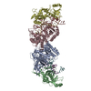 8jr8C M: map data used to model this data C: citing same article ( |
|---|---|
| Similar structure data |


-Links
 PDBj
PDBj


- Assembly
| Deposited unit | 
|
|---|---|
| 1 |
|
-Components
| #1: Protein | Mass: 58091.410 Da / Num. of mol.: 4 Source method: isolated from a genetically manipulated source Source: (gene. exp.) Maribacter polysiphoniae (bacteria) / Production host: #2: Protein | Mass: 53270.594 Da / Num. of mol.: 4 Source method: isolated from a genetically manipulated source Source: (gene. exp.) Maribacter polysiphoniae (bacteria) / Production host: #3: RNA chain | Mass: 6651.949 Da / Num. of mol.: 4 / Source method: obtained synthetically / Source: (synth.) Maribacter polysiphoniae (bacteria)#4: DNA chain | Mass: 7675.000 Da / Num. of mol.: 4 / Source method: obtained synthetically / Source: (synth.) Maribacter polysiphoniae (bacteria)Has protein modification | N | |
|---|
-Experimental details
-Experiment
| Experiment | Method: ELECTRON MICROSCOPY |
|---|---|
| EM experiment | Aggregation state: PARTICLE / 3D reconstruction method: single particle reconstruction |
- Sample preparation
| Component | Name: Maribacter polysiphoniae short Ago complexed with TIR-APAZ Type: COMPLEX / Entity ID: all / Source: RECOMBINANT |
|---|---|
| Source (natural) | Organism: Maribacter polysiphoniae (bacteria) |
| Source (recombinant) | Organism: |
| Buffer solution | pH: 7.5 |
| Specimen | Embedding applied: NO / Shadowing applied: NO / Staining applied: NO / Vitrification applied: YES |
| Vitrification | Cryogen name: ETHANE |
- Electron microscopy imaging
| Experimental equipment |  Model: Titan Krios / Image courtesy: FEI Company |
|---|---|
| Microscopy | Model: FEI TITAN KRIOS |
| Electron gun | Electron source:  FIELD EMISSION GUN / Accelerating voltage: 300 kV / Illumination mode: FLOOD BEAM FIELD EMISSION GUN / Accelerating voltage: 300 kV / Illumination mode: FLOOD BEAM |
| Electron lens | Mode: BRIGHT FIELD / Nominal defocus max: 2400 nm / Nominal defocus min: 1000 nm |
| Specimen holder | Cryogen: NITROGEN / Specimen holder model: FEI TITAN KRIOS AUTOGRID HOLDER |
| Image recording | Electron dose: 40 e/Å2 / Film or detector model: GATAN K2 SUMMIT (4k x 4k) |
- Processing
| EM software | Name: PHENIX / Category: model refinement | ||||||||||||||||||||||||
|---|---|---|---|---|---|---|---|---|---|---|---|---|---|---|---|---|---|---|---|---|---|---|---|---|---|
| CTF correction | Type: PHASE FLIPPING AND AMPLITUDE CORRECTION | ||||||||||||||||||||||||
| 3D reconstruction | Resolution: 3.3 Å / Resolution method: FSC 0.5 CUT-OFF / Num. of particles: 183347 / Symmetry type: POINT | ||||||||||||||||||||||||
| Refine LS restraints |
|
