 Movie
Movie Controller


+ Open data

- Basic information
| Entry | Database: PDB / ID: 8jr8 | ||||||||||||||||||||||||
|---|---|---|---|---|---|---|---|---|---|---|---|---|---|---|---|---|---|---|---|---|---|---|---|---|---|
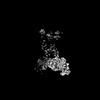
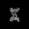
| Title | MapSPARTA dimer bound with guide-target | ||||||||||||||||||||||||
 Components Components |
| ||||||||||||||||||||||||
 Keywords Keywords | DNA BINDING PROTEIN/DNA/RNA / SPARTA / Ago / Tir / DNA BINDING PROTEIN-DNA-RNA complex | ||||||||||||||||||||||||
| Function / homology |  Function and homology information Function and homology information | ||||||||||||||||||||||||
| Biological species |  Maribacter polysiphoniae (bacteria) Maribacter polysiphoniae (bacteria) | ||||||||||||||||||||||||
| Method | ELECTRON MICROSCOPY / single particle reconstruction / cryo EM / Resolution: 3.48 Å | ||||||||||||||||||||||||
 Authors Authors | Huang, P.P. / Li, Z.X. / Guo, L.J. / Xiao, Y.B. / Chen, M.R. | ||||||||||||||||||||||||
| Funding support |  China, 5items China, 5items
| ||||||||||||||||||||||||
 Citation Citation | Journal: To Be Published Title: Cryo-EM Structure of MapSPARTA Authors: Huang, P.P. / Guo, L.J. / Li, Z.X. / Yan, P.R. / Chen, M.R. / Xiao, Y.B. | ||||||||||||||||||||||||
| History |
|
- Structure visualization
| Structure viewer | Molecule: MolmilJmol/JSmol |
|---|
- Downloads & links
-Download
| PDBx/mmCIF format | 8jr8.cif.gz | 341.4 KB | Display | PDBx/mmCIF format |
|---|---|---|---|---|
| PDB format | pdb8jr8.ent.gz | 266.8 KB | Display | PDB format |
| PDBx/mmJSON format | 8jr8.json.gz | Tree view | PDBx/mmJSON format | |
| Others |  Other downloads Other downloads |
-Validation report
| Arichive directory | https://data.pdbj.org/pub/pdb/validation_reports/jr/8jr8ftp://data.pdbj.org/pub/pdb/validation_reports/jr/8jr8 | HTTPS FTP |
|---|
-Related structure data
| Related structure data | 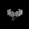 36592MC  8wp2C M: map data used to model this data C: citing same article ( |
|---|---|
| Similar structure data |


-Links
 PDBj
PDBj


- Assembly
| Deposited unit | 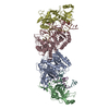
|
|---|---|
| 1 |
|
-Components
| #1: RNA chain | Mass: 6651.949 Da / Num. of mol.: 2 / Source method: obtained synthetically / Details: guide RNA / Source: (synth.) Maribacter polysiphoniae (bacteria)#2: DNA chain | Mass: 7675.000 Da / Num. of mol.: 2 / Source method: obtained synthetically / Details: target DNA / Source: (synth.) Maribacter polysiphoniae (bacteria)#3: Protein | Mass: 58091.410 Da / Num. of mol.: 2 Source method: isolated from a genetically manipulated source Source: (gene. exp.) Maribacter polysiphoniae (bacteria) / Gene: LX92_01809 / Production host: #4: Protein | Mass: 53270.594 Da / Num. of mol.: 2 Source method: isolated from a genetically manipulated source Source: (gene. exp.) Maribacter polysiphoniae (bacteria) / Gene: LX92_01810 / Production host: Has protein modification | N | |
|---|
-Experimental details
-Experiment
| Experiment | Method: ELECTRON MICROSCOPY |
|---|---|
| EM experiment | Aggregation state: PARTICLE / 3D reconstruction method: single particle reconstruction |
- Sample preparation
| Component | Name: Maribacter polysiphoniae short Ago complexed with TIR-APAZ Type: COMPLEX / Entity ID: all / Source: MULTIPLE SOURCES |
|---|---|
| Source (natural) | Organism: Maribacter polysiphoniae (bacteria) |
| Source (recombinant) | Organism: |
| Buffer solution | pH: 7.5 |
| Specimen | Embedding applied: NO / Shadowing applied: NO / Staining applied: NO / Vitrification applied: YES |
| Vitrification | Cryogen name: ETHANE |
- Electron microscopy imaging
| Experimental equipment |  Model: Titan Krios / Image courtesy: FEI Company |
|---|---|
| Microscopy | Model: FEI TITAN KRIOS |
| Electron gun | Electron source:  FIELD EMISSION GUN / Accelerating voltage: 300 kV / Illumination mode: FLOOD BEAM FIELD EMISSION GUN / Accelerating voltage: 300 kV / Illumination mode: FLOOD BEAM |
| Electron lens | Mode: BRIGHT FIELD / Nominal defocus max: 2400 nm / Nominal defocus min: 1000 nm |
| Specimen holder | Cryogen: NITROGEN / Specimen holder model: FEI TITAN KRIOS AUTOGRID HOLDER |
| Image recording | Electron dose: 40 e/Å2 / Detector mode: COUNTING / Film or detector model: GATAN K2 SUMMIT (4k x 4k) |
- Processing
| EM software | Name: PHENIX / Category: model refinement | ||||||||||||||||||||||||
|---|---|---|---|---|---|---|---|---|---|---|---|---|---|---|---|---|---|---|---|---|---|---|---|---|---|
| CTF correction | Type: PHASE FLIPPING AND AMPLITUDE CORRECTION | ||||||||||||||||||||||||
| 3D reconstruction | Resolution: 3.48 Å / Resolution method: FSC 0.143 CUT-OFF / Num. of particles: 79726 / Symmetry type: POINT | ||||||||||||||||||||||||
| Refine LS restraints |
|
