 Movie
Movie Controller

[English] 日本語
 Yorodumi
- PDB-8wme: Crystal Structure of Drosophila melanogaster D46A Dopamine N-Acet... -
+ Open data

- Basic information
| Entry | Database: PDB / ID: 8wme | ||||||
|---|---|---|---|---|---|---|---|
| Title | Crystal Structure of Drosophila melanogaster D46A Dopamine N-Acetyltransferase | ||||||
 Components Components | Arylalkylamine N-acetyltransferase 1 | ||||||
 Keywords Keywords | TRANSFERASE / Dopamine N-acetyltransferase(Dat) / GCN5-related N-acetyltransferase(GNAT) / Arylalkylamine N-acetyltransferase(AANAT) | ||||||
| Function / homology |  Function and homology information Function and homology informationchitin-based cuticle sclerotization / octopamine catabolic process / serotonin catabolic process / melatonin biosynthetic process / aralkylamine N-acetyltransferase / aralkylamine N-acetyltransferase activity / arylamine N-acetyltransferase activity / regulation of circadian sleep/wake cycle, sleep / catecholamine metabolic process / N-acetyltransferase activity ...chitin-based cuticle sclerotization / octopamine catabolic process / serotonin catabolic process / melatonin biosynthetic process / aralkylamine N-acetyltransferase / aralkylamine N-acetyltransferase activity / arylamine N-acetyltransferase activity / regulation of circadian sleep/wake cycle, sleep / catecholamine metabolic process / N-acetyltransferase activity / dopamine catabolic process / sleep / nucleus / cytoplasm Similarity search - Function | ||||||
| Biological species |  | ||||||
| Method |  X-RAY DIFFRACTION / SYNCHROTRON / MOLECULAR REPLACEMENT / Resolution: 2.283 Å X-RAY DIFFRACTION / SYNCHROTRON / MOLECULAR REPLACEMENT / Resolution: 2.283 Å | ||||||
 Authors Authors | Wu, C.Y. / Hu, I.C. / Cheng, H.C. / Lyu, P.C. | ||||||
| Funding support |  Taiwan, 1items Taiwan, 1items
| ||||||
 Citation Citation | Journal: To Be Published Title: Crystal Structure of Drosophila melanogaster D46A Dopamine N-Acetyltransferase in Complex with Acetyl-CoA Authors: Wu, C.Y. / Hu, I.C. / Cheng, H.C. / Lyu, P.C. | ||||||
| History |
|
- Structure visualization
| Structure viewer | Molecule: MolmilJmol/JSmol |
|---|
- Downloads & links
-Download
| PDBx/mmCIF format | 8wme.cif.gz | 99 KB | Display | PDBx/mmCIF format |
|---|---|---|---|---|
| PDB format | pdb8wme.ent.gz | 74.6 KB | Display | PDB format |
| PDBx/mmJSON format | 8wme.json.gz | Tree view | PDBx/mmJSON format | |
| Others |  Other downloads Other downloads |
-Validation report
| Arichive directory | https://data.pdbj.org/pub/pdb/validation_reports/wm/8wmeftp://data.pdbj.org/pub/pdb/validation_reports/wm/8wme | HTTPS FTP |
|---|
-Related structure data

-Links
 PDBj
PDBj

- Assembly
| Deposited unit | 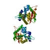
| ||||||||
|---|---|---|---|---|---|---|---|---|---|
| 1 | 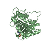
| ||||||||
| 2 | 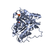
| ||||||||
| Unit cell |
|
-Components
| #1: Protein | Mass: 24400.057 Da / Num. of mol.: 2 / Mutation: D46A Source method: isolated from a genetically manipulated source Source: (gene. exp.) Production host:  References: UniProt: Q94521 #2: Chemical |   Mass: 238.305 Da / Num. of mol.: 2 / Source method: obtained synthetically / Formula: C8H18N2O4S / Comment: pH buffer*YM Mass: 238.305 Da / Num. of mol.: 2 / Source method: obtained synthetically / Formula: C8H18N2O4S / Comment: pH buffer*YM#3: Water | ChemComp-HOH / |  Mass: 18.015 Da / Num. of mol.: 146 / Source method: isolated from a natural source / Formula: H2O Mass: 18.015 Da / Num. of mol.: 146 / Source method: isolated from a natural source / Formula: H2OHas ligand of interest | N | |
|---|
-Experimental details
-Experiment
| Experiment | Method: X-RAY DIFFRACTION / Number of used crystals: 1 |
|---|
- Sample preparation
| Crystal | Density Matthews: 2.24 Å3/Da / Density % sol: 45.19 % |
|---|---|
| Crystal grow | Temperature: 277 K / Method: vapor diffusion, hanging drop / pH: 7 / Details: 1.0M NaH2PO4, 1.6M K2HPO4, 0.1M HEPES, 0.2M NaCl |
-Data collection
| Diffraction | Mean temperature: 100 K / Serial crystal experiment: N |
|---|---|
| Diffraction source | Source: SYNCHROTRON / Site: NSRRC / Beamline: BL13B1 / Wavelength: 0.99984 Å |
| Detector | Type: ADSC QUANTUM 315r / Detector: CCD / Date: Apr 14, 2021 |
| Radiation | Protocol: SINGLE WAVELENGTH / Monochromatic (M) / Laue (L): M / Scattering type: x-ray |
| Radiation wavelength | Wavelength: 0.99984 Å / Relative weight: 1 |
| Reflection | Resolution: 2.28→30 Å / Num. obs: 18954 / % possible obs: 96.1 % / Redundancy: 5.5 % / Biso Wilson estimate: 31.55 Å2 / Rmerge(I) obs: 0.116 / Rpim(I) all: 0.054 / Rrim(I) all: 0.128 / Χ2: 0.881 / Net I/σ(I): 13.46 |
- Processing
| Software |
| ||||||||||||||||||||||||||||||||||||||||||||||||
|---|---|---|---|---|---|---|---|---|---|---|---|---|---|---|---|---|---|---|---|---|---|---|---|---|---|---|---|---|---|---|---|---|---|---|---|---|---|---|---|---|---|---|---|---|---|---|---|---|---|
| Refinement | Method to determine structure: MOLECULAR REPLACEMENT / Resolution: 2.283→28.776 Å / SU ML: 0.28 / Cross valid method: THROUGHOUT / σ(F): 1.36 / Phase error: 25.32 / Stereochemistry target values: ML
| ||||||||||||||||||||||||||||||||||||||||||||||||
| Solvent computation | Shrinkage radii: 0.9 Å / VDW probe radii: 1.11 Å / Solvent model: FLAT BULK SOLVENT MODEL | ||||||||||||||||||||||||||||||||||||||||||||||||
| Displacement parameters | Biso max: 79.67 Å2 / Biso mean: 33.8521 Å2 / Biso min: 15.78 Å2 | ||||||||||||||||||||||||||||||||||||||||||||||||
| Refinement step | Cycle: final / Resolution: 2.283→28.776 Å
| ||||||||||||||||||||||||||||||||||||||||||||||||
| LS refinement shell | Refine-ID: X-RAY DIFFRACTION / Rfactor Rfree error: 0
|
